por
Lauren Dubinsky, Senior Reporter | December 16, 2015
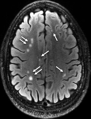
Multiple 'brain scars' in a man
with blast-related MTBI
Courtesy of RSNA
MR revealed brain damage in a large percentage of active duty military personnel who experienced blast-related mild traumatic brain injury (MTBI), according to a new study published online in the journal Radiology. This is the largest study ever conducted using advanced brain imaging on active military personnel.
"MTBI patients, by DOD criteria, are supposed to have normal routine imaging exams, so seeing abnormal exams in over 50 percent was surprising to us," Dr. Gerard Riedy from the National Intrepid Center of Excellence (NICoE) at the Walter Reed National Military Medical Center, told HCB News.
Physicians currently assess MTBI by evaluating the patients' behavior and recollection of events including post-traumatic amnesia and loss of consciousness. MR is not commonly used to assess these patients, but Riedy and his fellow researchers felt there was a need for a more definitive marker for MTBI.


Ad Statistics
Times Displayed: 352364
Times Visited: 21072 MIT labs, experts in Multi-Vendor component level repair of: MRI Coils, RF amplifiers, Gradient Amplifiers Contrast Media Injectors. System repairs, sub-assembly repairs, component level repairs, refurbish/calibrate. info@mitlabsusa.com/+1 (305) 470-8013
"MRI is expensive. Most mild TBI patients recover in three to six months, therefore it is not 'cost efficient' to scan everyone with TBI. These types of scans are typically reserved for complicated cases or for people that are not recovering as expected," wrote Riedy. "However, that fact, that they don't get MRIs, does not mean they don't have damage to the brain, we just have not imaged or documented that damage."
Riedy and his researchers used MR to evaluate 834 military personnel with MTBI related to blast injuries. A little more than 84 percent of the patients reported one or more blast-related incidents and 63 percent reported loss of consciousness when the injury occurred.
The MR scans showed the presence of white matter T2 hyperintensities, which are referred to as "brain scars", in 52 percent of the patients. It was a surprise to the researchers because it's expected that MTBI patients will have normal MR scans.
The MR exams that the researchers performed on the patients were not "routine MR" exams, but rather exams that were specially designed by their team of scientists to image patients with TBI.
The researchers also found that pituitary abnormalities were shown in almost a third of MTBI patients. The pituitary gland is located in the base of the brain and controls other endocrine gland functions.
They are currently building a database of advanced imaging data and hope to start to connect the data with the more subjective symptoms related to MTBI. For example, a scar on the brain scan is an objective finding and the researchers will use that to build a foundation for the right diagnosis of MTBI and then add in the subjective measures later.
Another area that they are focusing on is the diagnosis of post-traumatic stress disorder (PTSD), which is difficult to diagnose because the symptoms of PTSD and TBI are similar. In addition, the treatment for TBI is unlikely to work for PTSD and vice versa.
Riedy thinks that MR will become the standard of care for patients with MTBI in the future. "MRI is the best way to look for traumatic damage to the brain," he wrote. "When you have trauma to your knee, you get an X-ray or an MRI, when to land on your shoulder and have pain, you get an X-ray or an MRI, when an IED explodes and you get knocked unconscious you get…"


