por
Christina Hwang, Contributing Reporter | April 26, 2016

Allows researchers to view
biopsies with level of detail
similar to a microscope
Courtesy: University of Southampton
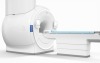
By using advanced 3-D X-ray imaging technology, new insights can be found into the way idiopathic pulmonary fibrosis (IPF), a disease that causes lung tissue to become thick, stiff or scarred, forms in the body.
Doctors and researchers from the University of Southampton, Royal Brompton Hospital, National Jewish Health and University College Dublin generated computer images using Microfocus CT to show active fibrosis areas, each identified by a different color.
The Microfocus CT can scan inside objects in great detail and was used to examine biopsy tissues from the lung by rotating 360 degrees while taking thousands of 2-D images, which were then combined to build 3-D images.


Ad Statistics
Times Displayed: 346858
Times Visited: 21062 MIT labs, experts in Multi-Vendor component level repair of: MRI Coils, RF amplifiers, Gradient Amplifiers Contrast Media Injectors. System repairs, sub-assembly repairs, component level repairs, refurbish/calibrate. info@mitlabsusa.com/+1 (305) 470-8013
“An important point is that the MicroCT technique can be performed on the same sample currently used only for 2-D microscopy analyses,” Dr. Mark Jones, lead author of the study, a Wellcome Trust fellow from the University of Southampton and University Hospital Southampton, told HCB News.
The researchers found that there were large numbers of individual sites of active disease scarring, which contradicts former studies that indicated active scarring in IPF progressed from the outside of the lung to the inside, the study reported.
Dr. Jones said that accurate diagnosis of IPF can be challenging due to the tools currently available in the market.
“Ultimately we hope that this technology will help us to improve both our understanding of how IPF develops, and how it is diagnosed,” said Dr. Jones. He added that this will help to ensure that the right patient is given a targeted treatment.
In the past few years, two drugs have been approved worldwide for the treatment of IPF, and Dr. Jones said both slow down the rate of lung scarring, which means that getting the correct diagnosis as early as possible is very important for each patient.
The Microfocus CT was originally designed for the analysis of engineering parts such as jet turbine blades, rather than lung tissue. “Designed as a particularly flexible, multi-function engineering system, we were able to develop and optimise the system configuration in a way that more standard machines would not normally allow,” Ian Sinclair, Director of the µ-VIS Centre for Computed Tomography, told HCB News.