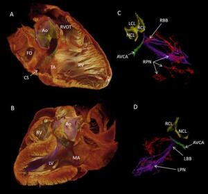
Scientists create first 3-D image of the heart’s cardiac conduction system
por John R. Fischer, Senior Reporter | August 15, 2017
Cardiology

3D image of cardiac conduction system
Heart surgeons may soon have a new tool that could prevent them from damaging tissue during heart surgery.
Scientists from Liverpool John Moores University, the University of Manchester, Aarhus University and Newcastle University have created the first 3-D image of the cardiac conduction system, through the use of contrast-enhanced microcomputed tomography, compiling their findings in a study published in Scientific Reports.
The study’s authors argue that their depiction of the system is more accurate, compared to two-dimensional computer imaging and textbook representations, and could act as a guide for surgeons during heart surgery, to prevent them from accidentally damaging tissue.
“Most of this delicate system is buried within the heart muscle and is not visible during heart surgeries,” Dr. Halina Drobrzynski, a senior lecturer in cardiac biology at the University of Manchester and one of the authors of the study, told HCB News. “So it can be damaged, for example, during aortic valve replacements. Therefore, we hope that our model can help heart doctors to understand better the location of different components of this electrical system within the heart, especially because we have presented the data as a 3-D video.”
The microCT technology is already used to depict images of other organs, include the lungs, liver and kidney. The images act as a guide for treating various ailments, including fibrosis of the lungs, kidney stones or corrosion of the liver vasculature in organs removed during transplants.
Using the 3-D image of the cardiac conduction system, Drobrzynski says doctors can be guided to better address and treat cardiac arrhythmia and other heart conditions.
“The 3-D representation method could also be used to make postmortem investigations in congenital heart defects, identifying scar regions after heart attacks, showing changes in heart structures in disease states like enlarged hearts due to high blood pressure, or narrowing of the heart valves,” said Drobrzynski.
Drobrzynski says that the 3-D imaging technology requires more research but could be used more to create strong guides for better understanding the inner workings of the heart.
“It is too early to say if this technology will become a standard tool for guiding heart doctors and surgeons in the future,” she said. “But it will, for sure, provide them with an educational tool to visualize in great detail the complex anatomy of the heart and thus, better understand the normal function of the cardiac conduction system and disorders of heart rhythms.”
Back to HCB News
1(current)
You Must Be Logged In To Post A CommentRegistroRegistrarse es Gratis y Fácil. Disfruta de los beneficios del Mercado de Equipos Médicos Nuevos y Usados líder en el mundo. ¡Regístrate ahora! |
|


