Ultrasound technique can explore why fat cells put on fat
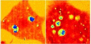
Light is usually used to see inside live cells, but researchers at the University of Nottingham in England developed a new technique that uses sound. The ultrasound technique can potentially be used for cancer diagnosis and stem-cell transplants.
The technique uses shorter-than-optical wavelengths of sound and provides information about the structure, mechanical properties and behavior of living cells. This may compete with the optical super-resolution techniques that won the Nobel Prize for Chemistry in 2014.
“People are most familiar with ultrasound as a way of looking inside the body — in the simplest terms, we’ve engineered it to the point where it can look inside an individual cell,” Matt Clark, professor at the university, said in a statement. “Nottingham is currently the only place in the world with this capability.”
Conventional optical microscopy uses light, and the size of the smallest object you can see is limited by the wavelength. The fluorescent dyes that optical super-resolution imaging uses are usually toxic and it takes a lot of time and light to observe and reconstruct an image, which is damaging to cells.
Sound doesn’t have a high-energy payload like light does. Because of that, the researchers were able to use smaller wavelengths to see tinier things and get to higher resolutions without damaging the cell biology.
“A great thing is that, like ultrasound on the body, ultrasound in the cells causes no damage and requires no toxic chemicals to work,” said Clark. “Because of this we can see inside cells that one day might be put back into the body, for instance as stem cell transplants.”