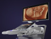
Mammography has been the standard for screening breast cancer for many decades, but its drawbacks are leading the industry to pursue better options. A research team at Eindhoven University of Technology in the Netherlands may have found one.
Dynamic Contrast Specific Ultrasound Tomography identifies cancerous tumors by evaluating the structure of blood vessels. Unlike mammography, which tightly squeezes the breast between two plates, this technology requires the patient to lie on a table as their breast is suspended in a bowl.
This technology builds on the prostate cancer detection method that the university developed in which the physician injects the patient with microbubbles. Those microbubbles are then monitored as they travel through the blood vessels in the prostate.
The formation of chaotic microvessels is an indicator that cancer is growing, so this method can be used to pinpoint the location of the tumor. It has proven to work well for diagnosing prostate cancer and is being tested in hospitals in the Netherlands and China, and will soon be tested in Germany.
However, there have been complications in translating those benefits to breast imaging, so the researchers made a few adjustments to it. The microbubbles vibrate in the blood at the same rate as the sound produced by the echoscanner and even twice that, which is referred to as the second harmonic.
The researchers discovered that the second harmonic was slightly delayed by the gas bubbles. But when the delay is measured, they can localize the air bubbles without causing any disturbance because the harmonic generated by the body tissue isn’t delayed.
That difference can only be seen if the sound is captured on the other side, which makes the approach ideal for organs that can be approached from two sides, such as the breast.
The researchers are now working on bringing together an international medical team to start conducting preclinical studies. But it will be another ten years or so until the technology will be ready for clinical use and it will likely be used in combination with other methods.
A proof of concept discussing the technology was published earlier this month in the online journal, Scientific Report.