por
Lauren Dubinsky, Senior Reporter | December 11, 2015
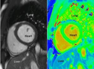
T1-maps on right
Courtesy of Dr. Alexander Liu
Researchers at Oxford University created a technique that shows healthy and diseased heart tissue in greater detail on MR images without the use of contrast agents.
The researchers used the property of hydrogen atoms to create a pixel-by-pixel map of the heart called a T1-map. The name of the mapping technique is ShMOLLI.
Stress scans of the heart that use MR imaging require contrast agents, such as Gadolinium, to highlight areas of the heart with decreased blood flow and Adenosine, which causes effects similar to exercise.


Ad Statistics
Times Displayed: 14289
Times Visited: 25 Final days to save an extra 10% on Imaging, Ultrasound, and Biomed parts web prices.* Unlimited use now through September 30 with code AANIV10 (*certain restrictions apply)
Patients with severe kidney failure who usually have a higher risk of heart disease can't clear Gadolinium, so they often cannot undergo these MR exams. But the T1 maps hold promise for this patient population.
"T1 mapping allows us to look in finer detail at the heart in a non-invasive way, which has not been possible before," Dr. Stefan Piechnik, one of the researchers, said in a statement. "We can now get results without Gadolinium, meaning we have a technique that is safer and quicker and can be used with more people."
With traditional MR images, there is a dependency on the physicians' interpretation, which can vary, but the T1 map gives them an objective number that can be coded in colors, which may be more subjective.
The T1 map works by measuring T1 values across the heart to determine the exact location of the problem. It takes around three minutes to map the entire heart and the values are turned into a color map.
The T1 map is not only useful for evaluating conditions in the heart — it can be used to identify unhealthy tissue in other parts of the body. Every type of tissue in the body has a range of normal T1 values and any value outside of that range might indicate disease.
The researchers are currently applying the technique to other organs. The U.K. Biobank, which is a large long-term biobank study, has a goal to image 100,000 patients in the U.K. using the technique.
They are hoping to translate the technique into clinical use within the next five years.