por
Lauren Dubinsky, Senior Reporter | December 30, 2014
To diagnose Alzheimer's physicians are forced to mainly rely on clinical evidence of mental decline, by which time the disease has already caused severe brain damage.
But that may soon change thanks to breakthrough research by a team of scientists and engineers from Northwestern University. They have developed a new non-invasive MR method that can detect the condition in an early stage.
And the probe actually improved memory in test animals, according to the study.


Ad Statistics
Times Displayed: 194156
Times Visited: 5654 For those who need to move fast and expand clinical capabilities -- and would love new equipment -- the uCT 550 Advance offers a new fully configured 80-slice CT in up to 2 weeks with routine maintenance and parts and Software Upgrades for Life™ included.
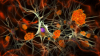
The team created a probe that combines a magnetic nanostructure and an antibody that targets the amyloid beta brain toxins, which are known culprits behind the disease. When an MR exam is performed, the toxins appear as dark areas in the images.
"The really big benefit is being able to monitor the level of the toxins that cause memory loss and dementia — right now it's not possible," William L. Klein, lead researcher and neuroscientist at the university, told DOTmed News. "The biggest immediate benefit would be for testing how well investigational new drugs actually work."
A paper on the team's research, "Towards non-invasive diagnostic imaging of early-stage Alzheimer's disease," was recently published by the journal Nature Nanotechnology. Klein said they have a prototype of the probe for use in humans, but that "there is plenty of work to do before this new probe reaches the clinic."
Despite a massive effort to find effective medications to combat Alzheimer's, there has been no significant success to date. But using amyloid beta as a biomarker may prove key to finding an effective drug.
Even though memory improvement was not the focus of the team's research, they found that the probe improved memory because it binds to the toxins and prevents them from doing further damage.
Conventional technology detects plaques, instead of amyloid beta toxins, which occur when therapeutic intervention would be very late and less successful. The mobile amyloid beta toxins attack the synapses of neurons, which destroys memory and causes the neurons to die. Over time the amyloid beta accumulates and begins to stick together, which then turns into amyloid plaque.
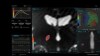
In an experiment, the MR probe was administered intravenously to mice with Alzheimer's disease and healthy control mice. The team found that the toxins were dark in the hippocampus in brain MR scans for the mice with Alzheimer's, but not in the control group.
The team also used the MR probe on human brain tissue from patients who died from Alzheimer's and compared that tissue with samples from those who did not have the disease. They found that administering the MR probe allowed them to see the dark areas in the brains of the patients with Alzheimer's.
Back to HCB News