por
Lauren Dubinsky, Senior Reporter | April 20, 2018
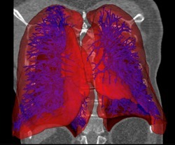
AI organ segmentation of lungs
and vasculature
Aether will soon launch its new Artificial Intelligence-powered medical imaging software to take its 3-D organ printing technology to the next level.
“Presently there are many challenges in the 3-D bioprinting field,” Ryan Franks, CEO of Aether, told HCB News. “I believe many of these challenges can be solved by AI.”
Researchers struggle to achieve the correct line width for extruded materials, calculate the precision position of multiple syringe needles and find and correct errors during and after a print.


Ad Statistics
Times Displayed: 14290
Times Visited: 25 Final days to save an extra 10% on Imaging, Ultrasound, and Biomed parts web prices.* Unlimited use now through September 30 with code AANIV10 (*certain restrictions apply)
“By using cameras to analyze the printing process from initial calibration all the way to completed print, machine vision, deep learning and other AI techniques can completely revolutionize the entire process from start to finish,” Ryan Franks, CEO of Aether, told HCB News.
The new AI software is capable of performing Automatic Segmentation and Reconstruction (ASAR). It leverages adaptable deep learning models and a variety of AI and image processing techniques to enable users to segment organs and tissues and reconstruct them as digital 3-D models.
Those models can be used along with organ fabrication systems to create exact replicas of patient anatomy made from ultra-realistic synthetic human tissue, bone, fat, vasculature and blood. Surgeons can use those replicas to practice upcoming procedures.
Other software on the market can take multiple hours to create those models, but ASAR can do it in a matter of minutes or seconds. In addition, it’s completely automatic and requires no editing tools, calibration or human intervention.
The software currently uses CT scan datasets to create the 3-D models, but Aether has plans to extend this across all major modalities including MR, X-ray and angiography.
“We're really excited about converting CT scans directly to multi material 3-D files — it’s completely revolutionary for surgical training and medical image visualization,” said Franks. “However, surgical training is just a stop on the road to true implantable organs.”
Aether is working with labs at universities like Harvard Medical School (HMS) to make 3-D-printed viable organs for transplantation a possibility. The Jang Laboratory at HMS plans to modify Aether’s multi-material synthetic organ model 3-D printing process to print organs with real human cells.
“Imagine transplants not of generic organs, but a detailed duplicate of the orginal, with shape and structure replicated directly from a scan of the patient,” said Franks. “We are creating the technology that the most brilliant minds will use to make personalized organs on demand a reality.”
Back to HCB News