por
John R. Fischer, Senior Reporter | November 01, 2017
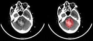
ParallelDots has developed AI software
for detecting brain hemorrhages on
CT scans
Radiologists may soon have a new point of reference for confirming the presence of brain bleeds in patients.
ParallelDots Inc., a U.S. and India-based company that develops real-world artificial intelligence applications, has created an AI software solution, RADnet, for detecting brain hemorrhages on CT scans to serve as a second opinion for radiologists and decrease diagnosis time, in an aim to increase survival rates. Its research findings were published in a
research paper.
“It's important for the radiologist to know about a critical case as soon as it comes,” Angam Parashar, a co-founder of ParallelDots, told HCB News. “It allows them to save time and provide faster diagnosis to the patient. We see radiologists using our technology for second opinions. Second opinions are very much needed in many cases in radiology. AI can help radiologists in making accidental findings in medical images.”


Ad Statistics
Times Displayed: 18554
Times Visited: 52 Brand-New FDA-cleared Advanced Ultrasound Medical Device available for sale or lease to Wound Care Centers or any other Medical Facilities.The Arobella 1000D is designed for non-contact or debridement ultrasound wound healing therapy, or any other wounds
Traditional approaches for detecting hemorrhages are time consuming, requiring a radiologist to be present at every moment to make visual inspections of conditions and manual quantitative estimations of the size of the hematoma and midline shift.
The company developed the software using deep neural networks and obtained anonymous medical data in the form of 2-D CT slice sequences from hospitals. Medical professionals then annotated the data to prepare it for machine learning, marking areas with signs of brain hemorrhages, if present, with loose approximate boundaries.
A convolutional neural network, specifically DenseNet, then modeled each 2-D slice of the CT scan images. The network was made to focus its attention toward hemorrhage regions in each 2-D slice and a bidirectional long short-term memory (LSTM) network was used to model the 3-D aspect of a CT scan.
Company researchers compared RADnet’s performance to that of three senior radiologists, testing its accuracy, recall, precision and F1 score on 77 brain CTs. The software scored a higher recall score than two of the three and the same one as the other. It also showed the same degree of accuracy as one and a higher F1 score than two.
Parashar warns that the software is not meant to replace radiologists but to assist in their workflow, and that further testing is necessary.
“One big challenge that we see is around the performance of the AI system,” he said. “We are dealing with critical cases here. The AI technology needs to be really accurate to be meaningful to a radiologist. Additionally, it is important to consider other clinical parameters when developing the AI system. Combining all the information with the medical imaging data is another challenge that needs to be addressed.”
The AI model was trained on 205 CT scans only, and it is only with this limited amount of data that researchers were able to match the accuracy of the radiologists.
The paper will soon be submitted to peer-reviewed journals.


