por
Lauren Dubinsky, Senior Reporter | October 20, 2017
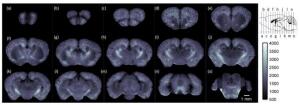
Using cryo-MOST, the researchers
acquired a brain-wide map of an
Alzheimer mouse model showing
that senile plaques
had spread to the entire brain
Researchers in China developed a new imaging system that has the potential to accelerate drug development for Alzheimer's disease.
Cryo-micro-optical sectioning tomography is a better alternative for monitoring brain changes associated with the disease in mouse models, according to a study published in
Optics Letters.
"We tried to observe the redox state of Alzheimer's in mouse brain [models] and found some interesting new phenomena," Jing Yuan, one of the researchers, told HCB News. "[We wondered] if there was any other useful autofluorescence we could find from tissue and then began this new project."


Ad Statistics
Times Displayed: 14290
Times Visited: 25 Final days to save an extra 10% on Imaging, Ultrasound, and Biomed parts web prices.* Unlimited use now through September 30 with code AANIV10 (*certain restrictions apply)
Mice are valuable for studying the biology of Alzheimer's and testing new drugs because proteins called senile plaques accumulate in both human and mice brains. The cryo-MOST system was designed to better image those proteins in the whole mouse brain.
Since it's optical, the cryo-MOST system generates more detailed information than other imaging modalities such as MR and PET. It also doesn't require external dyes or labels because it takes advantage of the natural fluorescence of senile plaques after exposure to excitation light.
For the study, Yuan and his team used cryo-MOST to image the whole brain of a 17-month mouse with Alzheimer's. The images revealed that senile plaques had spread to the whole brain.
That indicates that the disease doesn't only affect memory and intelligence, but might also lead to the deterioration of other brain functions.
In the case of humans, the cryo-MOST system can only be used to study deceased donors since the size of the tissue imaged is limited by the maximum moving range of the mechanical stage. It may also be useful for visualizing other biological molecules that exhibit autofluorescence in the kidney and liver.
The research team is currently working to improve the system's automation in order to accelerate image acquisition, and making enhancements that will improve image quality. That will enable the system to be used for more applications.
They are also continuing their research into studying senile plaque distribution and morphology changes as a result of Alzheimer's progression.
When asked if cryo-MOST will eventually become the standard for studying Alzheimer's, Yuan said that it has potential.
"Taking advantage of simple sample preparation, intrinsic autofluorescence, whole-brain imaging range and micron-level resolution, cryo-MOST [could] potentially become a useful research tool and standard for studying Alzheimer's," he added.
Back to HCB News