Para brindar el contenido más relevante a los lectores de HealthCare Business News, le pedimos que comparta un poco de información sobre quién es usted (tarda dos segundos y listo).
Registro
PET imaging used for the first time to evaluate Zika virus in mouse model
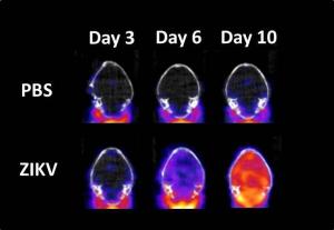
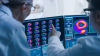
Bottom row: PET/CT mouse brain images at days 3, 6 and 10 post-infection with Zika virus Top row: PBS control mouse brain images Red area indicates neuroinflammation
A team at the U.S. Army Medical Research Institute of Infectious Diseases recently became the first to use PET imaging to evaluate brain inflammation in mice infected with the Zika virus.
“The results of the current study demonstrate that global neuroinflammation plays a significant role in the progression of the [Zika virus], and that [18F]DPA-714 PET imaging is a sensitive tool relative to histology for the detection of neuroinflammation,” Thomas M. Bocan, head of the in vivo imaging group at USAMRIID, told HCB News.
The traditional method for tracking the progression of infectious diseases is serial necropsies. The organ of interest is removed and a histological assessment is performed to define the pathology such as immunohistochemical staining for brain neuroinflammation.
“The methods are inherently variable because the disease does not proceed at the same pace in all animals,” said Bocan. “Since the analysis procedures are terminal, each animal can only be evaluated once, so larger numbers of animals are used to control for the variability in response.”
Using [18F]DPA-714 PET imaging, Bocan and his team found that levels of the Zika virus in the mouse brain increased from day three to day 10, post-infection. In that time frame, the mice also experienced a two- to six-fold increase in global brain neuroinflammation.
“[18F]DPA-714 PET imaging may be useful in dynamically characterizing the pathology associated with neurotropic viruses and the evaluation of therapeutics being developed for treatment of infectious diseases,” Bocan concluded.