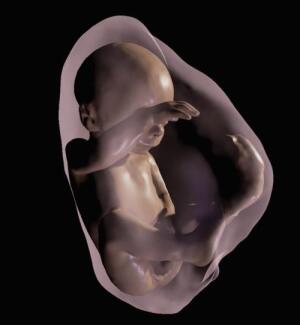
Ultrasound technology has greatly improved in recent years to provide parents with a better view of their unborn babies. Researchers are taking that one step further by leveraging a new technology called 3-D immersive visualization to transform ultrasound and MR data into a 3-D, virtual reality model of the baby.
The research will be presented next week at the annual meeting of the Radiological Society of North America in Chicago.
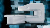
The researchers used sequentially-mounted MR slices to begin constructing the 3-D model. MR provides high-resolution fetal and placental imaging and is usually used during fetal evaluations when ultrasound can't provide adequate images.
The physician then selects the body parts that they want to be reconstructed in 3-D. Once an accurate 3-D model that includes the womb, umbilical cord, placenta and fetus is created, the virtual reality device can be programmed to incorporate the model.
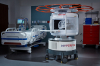
To create the virtual reality device, the researchers used the Oculus Rift 2 headset. This headset brings the users into an immersive environment in which they can hear heartbeat sounds and study the 3-D fetal anatomy by moving their head.
The virtual reality detail 3-D models are similar to the postnatal appearance of the newborn baby. The technology can recreate all of the internal structures of the fetus including the respiratory tract, which can help physicians evaluate abnormalities.
One of the technology's potential applications is to assess the state of the fetus' airways. If the ultrasound shows an abnormal mass near the fetal airway, the physician can use the 3-D images and headset to examine the entire length of the airway and make better informed decisions about delivery.
The technology can also be used to help coordinate care between multidisciplinary teams and provide the parents with better visual information so they can understand malformations and treatment decisions.
So far, the researchers have used this technology on patients at a clinic in Rio de Janeiro, which included a case in which the fetus had an abnormality that required postnatal surgery. They are hoping to use the technology on more patients over the next year.