UCSF develops 3-D virtual reality colonoscopy
por
Lauren Dubinsky, Senior Reporter | July 11, 2016
Courtesy of Noah Berger
Colonoscopies are invasive and often an unpleasant experience, but researchers at the University of California San Francisco (UCSF) have developed a non-invasive screening method that uses CT scans to generate a 3-D holograms of the patient's colon.
When patients are screened for breast, lung and prostate cancer, the clinicians are usually looking for the cancer itself, but in the case of colorectal cancer it would be too late. They can find a polyp before it becomes cancerous, but more patients need to get screened in order to have a significant impact on preventing the disease.
During a colonoscopy, the patient is under anesthesia and a six-foot-long flexible tube with a scope is inserted into the colon. The procedure takes 20 to 30 minutes and there is a risk of perforation, bleeding or infection.
The upside is that it's covered by Medicare and if polyps are detected, they can often be removed during screening because the patient is already sedated. But despite that, fewer than half of people who should get screened actually do.
CT colonography (CTC) uses thin slices of a CT scan to make the image appear 3-D on a flat screen. It can also morph into video views, which enhances the polyps, lesions and other precancerous anomalies.
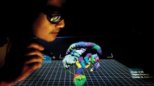
The UCSF researchers took it one step further with the addition of holograms. The clinicians wear 3-D glasses and as they move their head, a computer monitor with stereoscopic optical technology tracks the glasses and constructs a virtual holographic object that recreates the size and shape of the patient's anatomy.
The clinicians also use a stylus, which allows them to select the part of the scan they want to examine in more detail and to interact with it in a 3-D space. Essentially, they are able to eliminate the parts they don't want to see and improve what they do want to see.
"It’s a more engaging way to read large data sets," Dr. Judy Yee, professor and vice chair of the department of radiology and biomedical imaging at UCSF, said in a statement. "With the added dimension, you can see flat, more dangerous lesions better.”
This new method also holds promise for neurological, cardiac and musculoskeletal applications. Yee is hopeful that this can help demonstrate what radiology can bring to patient diagnosis and management for all parts of the body.
Yee and her colleagues are working to convince Medicare to provide reimbursement for CTC like many private insurers do. A CTC exam costs much less than conventional colonoscopy, but many Medicare patients still may not be able to pay for it out of pocket.
Yee's first study on virtual holographic CTC is underway, has already loaded 300 CTC scans, and the radiologists are being trained. UCSF also partnered with a Swedish company called Phase Holographic Imaging to accelerate skin cancer research.
|
|
|
You Must Be Logged In To Post A Comment
|