por
Lauren Dubinsky, Senior Reporter | January 12, 2016
Researchers at the University of Colorado Cancer Center have developed a new method for identifying and sparing healthy lung tissue during lung cancer radiotherapy without the need for additional testing. The National Institutes of Health funded a clinical trial that’s investigating the method and will be completed in three years.
“If our study is successful, we hypothesize that this new treatment approach to include functional imaging would reduce radiation-related side effects for patients that have undergone radiation therapy for lung cancer,” Yevgeniy Vinogradskiy, CU Cancer Center investigator and assistant professor in the Department of Radiation Oncology at the CU School of Medicine, told HCB News.
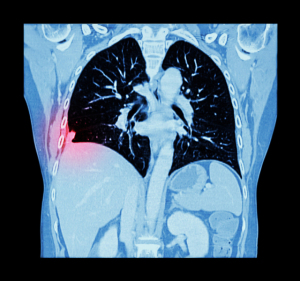
In the past, clinicians treated the lungs as a homogenous organ, but now they have realized that only certain areas should be treated and other areas should be spared. The idea behind the new method is to show the clinicians what areas of the lung should be spared from radiation.


Ad Statistics
Times Displayed: 14289
Times Visited: 25 Final days to save an extra 10% on Imaging, Ultrasound, and Biomed parts web prices.* Unlimited use now through September 30 with code AANIV10 (*certain restrictions apply)
The challenge is that lung tumors move along with the patient’s breathing, which makes any single image of the tumor’s position inaccurate for most of the breathing cycle. Previously, clinicians used rough estimates to account for the patient’s breathing motion during therapy.
When 4-D CT came into the picture in the early 2000s, it was a more efficient way to solve that problem. The technology uses a series of images shot over time to capture the position of the lung and tumor during all the phases of the breathing cycle.
Today, most lung cancer patients undergo 4-D CT exams so that radiation oncologists can develop a personalized treatment plan that takes their breathing motion into consideration. However, the limitation of 4-D CT is that it does not show the function of the surrounding lung tissue.
The new method combines air movement information from existing 4-D CT data with some equations to calculate lung function in the tissue that surrounds the tumor. Clinicians can then take that information and use advanced radiation delivery techniques to spare the part of the lungs used for breathing — thereby yielding significantly better outcomes.
The benefits are obvious for patients, but hospitals may also benefit from this new method.
“One of the neat things about the functional imaging approach we are taking is that the functional information is acquired using imaging that the patient receives as part of standard-of-care; therefore,no additional imaging procedure is required for the patient, which is convenient and saves both the hospital and the patient money,” wrote Vinogradskiy.
Back to HCB News