Philips' new HeartModel tool for the EPIQ 7 ultrasound
Collecting and analyzing heart measurements is traditionally a time-consuming, difficult process that results in variability. To address that, Royal Philips launched its new HeartModel tool for its EPIQ 7 ultrasound last Friday.
Conventional cardiac ultrasound technology takes up to three hours to perform 50 studies. “That’s a lot of time to spend doing measurements,” Dr. Wendy Tsang, cardiologist at Toronto General Hospital, told DOTmed News. “With the HeartModel, we are able to cut that down by almost two-thirds.”
In this new health care environment, health systems are searching for technology that can most efficiently and effectively provide them with an accurate diagnosis. In addition, cardiologists are under pressure to perform more studies in a day.
“There are so many uses for echocardiography right now and the volumes have been increasing and increasing,” said Tsang. “But it’s not like hospitals are hiring more physicians because they’re trying to maintain cost.”
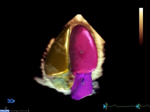
Cardiac chamber quantification from 3-D transthoracic echocardiography (3D TTE) has been shown to be superior to measurements obtained from 2-D studies but integrating it into routine clinical practice has been time-consuming. However, with EPIQ that process is automated.
“By being able to [use EPIQ], we can maintain a high standard of care for the patients, get more accurate results, more reproducible results and still keep our sanity because there are only so many hours in a day,” said Tsang.
In a recent study, Tsang and her fellow researchers used HeartModel to perform 3D TTE on 114 patients to assess the feasibility and accuracy of a fully automated algorithm that quantifies left atrial (LA) and left ventricular volumes (LV) and ejection fractions (EFs) without human interaction.
They found that automated quantification of LA and LV volumes and EFs is feasible in the majority of the patients and does not require specialized 3D TTE training. They concluded that this technique will improve integration of 3D TTE into routine clinical practice.
“This is the future of the way 3-D is going to be integrated into clinical practice,” said Tsang. “One of the biggest stumbling blocks is letting people use 3-D every day. This is a way that lets that happen and it’s going to make things better for patients."