por
Lauren Dubinsky, Senior Reporter | May 12, 2015
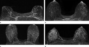
Axial postcontrast maximum intensity
projection MR images show (a) minimal,
(b) mild, (c) moderate, and (d) marked BPE
Courtesy of RSNA
As the health industry moves toward a more personalized approach to breast cancer screening, new research published in Radiology suggests MRI could potentially play a bigger part in that process than previously thought.
"To date, it's been difficult to assess the future risk of breast cancer for women, so there is a strong desire in the oncology community to identify ways to better determine this risk," Dr. Habib Rahbar, co-author of the study and a breast imaging expert at Seattle Cancer Care Alliance and assistant professor at the University of Washington, said in a statement.
Contrast-enhanced MRI is already used as a supplemental screening tool to mammography for women at high-risk. In fact, the American Cancer Society’s guidelines state that women with a 20 percent or higher lifetime chance of developing breast cancer should undergo both breast MRI and mammography screening every year.


Ad Statistics
Times Displayed: 194156
Times Visited: 5654 For those who need to move fast and expand clinical capabilities -- and would love new equipment -- the uCT 550 Advance offers a new fully configured 80-slice CT in up to 2 weeks with routine maintenance and parts and Software Upgrades for Life™ included.
For their study, Rahbar and his fellow researchers compiled breast MRI images from screenings conducted between January 2006 and December 2011. They looked specifically at high-risk patients ages 18 and over, with no history of breast cancer. They then evaluated the images for breast density and normal background parenchymal enhancement (BPE), which is when the background breast tissue shows up white on the MRI images.
The study found that the women with elevated amounts of BPE had a nine times higher chance of being diagnosed with breast cancer during the follow-up period compared to the women with low amounts of BPE, or none at all.
The researchers also found that mammographic breast density has no significant connection to cancer risk in this particular study. But BPE, on the other hand, may help physicians personalize screening and tailor treatment to each individual patient.
Previous studies have shown that dense breast tissue may put a woman at a higher risk of developing breast cancer, but Rahbar and his team are uncertain that including dense breast tissue as a risk factor can improve upon current risk assessment methods.
Women at high risk of developing breast cancer have the choice of undergoing supplemental screening mammography with MRI, therapy that blocks estrogen activity in the breast or a mastectomy. BPE may now help those women choose the most effective option.
Going forward, the researchers are conducting a larger study in order to validate their findings. They speculate that BPE is associated with inflammation in the early stages of a disease but they want to prove exactly why BPE might be a biomarker for breast cancer risk.
Back to HCB News