por
Lauren Dubinsky, Senior Reporter | June 11, 2014
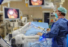
A recent study published online in the journal Pituitary found that intraoperative MRI (iMRI) paired with endoscopy for transsphenoidal surgery removes more pituitary tumor than conventional microscopy without iMRI. To date it's the largest published study to investigate the use of both iMRI and endoscopy.
Surgery is one of the main ways that pituitary tumors are treated but for patients with more difficult or larger tumors, even with the best efforts, the tumors may grow back. "We know that there is some limit to the best practices of surgery," Dr. Michael Chicoine, one of the study authors and associate professor of neurosurgery at the Washington University School of Medicine, told DOTmed News.
The iMRI gives the surgeons high-resolution images that show the areas of the tumor that remain and are not immediately apparent with an endoscope.


Ad Statistics
Times Displayed: 14289
Times Visited: 25 Final days to save an extra 10% on Imaging, Ultrasound, and Biomed parts web prices.* Unlimited use now through September 30 with code AANIV10 (*certain restrictions apply)
The study compared 446 patients at Barnes-Jewish Hospital in St. Louis and found that iMRI revealed that 56 out of 156 patients had incomplete tumor removal. For 15 of them, the additional tumor removal greatly improved their survival rate.
iMRI has been around for 10 to 15 years but it's constantly improving, said Chicoine. "This idea of wedding the endoscope with the intraoperative MRI is something that people have been dabbling with for years but our study was really the first to analyze a large population of patients with whom this strategy has been used and compare it to patients with whom this technology was not used," he added.
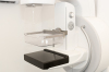
The study was performed using IMRIS' VISIUS Surgical Theatre and the surgeons had on-demand access to real-time data and imaging during the procedure from the operating room table. The surgeons didn't have to transport the patients since the VISIUS iMRI can move to the patients on ceiling-mounted rails.
Even though iMRI has been shown to provide benefits during surgery, it also comes along with a few challenges, one of them being safety.
"You have this six-ton very powerful magnet in the operating room and every metal object can potentially become a projectile in that environment," said Chicoine. But they were able to come up with a protocol to make sure all of the metal objects are outside of the high magnetic field areas.
Going forward, Chicoine and his fellow researchers are going to continue to work with MRI physicists and radiologists to try to come up with the best MR sequences in order to give the greatest amount of information in the most time efficient fashion during surgery.
"It's a fairly complicated scenario to use all of this technology — endoscopy, intraoperative MRI — and so we're continuing to streamline it and make it as efficient and effective as possible," said Chicoine.