por
Brendon Nafziger, DOTmed News Associate Editor | September 14, 2010
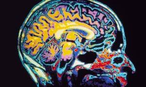
MRI scans of children and teens might be able to help doctors determine if their brains are developing normally, potentially helping physicians catch development problems before symptoms show up, according to researchers.
Using sophisticated mathematical tools, researchers at the Washington University School of Medicine in St. Louis could turn 5-minute scans of the brain into developmental assessments.
The work, published last week in Science, is based on studies from last year showing that MRI scans can reveal different organizational structures between kids and adults. In short, as brains mature, they start to rely on more distantly connected neural networks, instead of just tightly local ones.


Ad Statistics
Times Displayed: 101608
Times Visited: 6232 MIT labs, experts in Multi-Vendor component level repair of: MRI Coils, RF amplifiers, Gradient Amplifiers Contrast Media Injectors. System repairs, sub-assembly repairs, component level repairs, refurbish/calibrate. info@mitlabsusa.com/+1 (305) 470-8013
To test the subjects, scientists use a resting state functional connectivity scan coupled with a mathematical tool called a support vector machine. The support vector machine, a statistical learning system, is used in character recognition software and computational biology.
"It's a way that mathematicians have developed for predicting something with high specificity and sensitivity when you have huge amounts of data instead of one really good measurement," lead author Dr. Nico Dosenbach, a pediatric neurology resident at St. Louis Children's Hospital, said in prepared remarks. "Any one of these measurements doesn't tell you much, but if you put them together and use the right math to sift through and restructure them, you can get good predictive results."
For the experiment, the researchers used scans from around 238 subjects with ages ranging from seven to 30. Then they used about 200 of the scans to create an index of maturity. The researchers were able to predict, based on the scans, a subject's age with a high degree of accuracy.
The technique is about 91 to 92 percent accurate for comparing adults, from 25-29 years old, to children 7-11 years old, Dosenbach told DOTmed News by e-mail.
Still, it's too early to say if the technique will work the way the researchers hope - by letting doctors gauge the difference between normal and abnormal development. All the subjects in the current study were normal, and the researchers are only beginning to test if differences emerge using the technique on abnormal patients.
"We're currently working on that and the results seem very promising, but because of low sample sizes these results aren't ready for prime time, yet," Dosenbach said.
The researchers caution that the technique is probably too cost-prohibitive to be routinely used to screen children, as MRI scans can cost around $2,000 each, according to some estimates. However, many children with development problems often receive MRI scans, and the technique wouldn't really increase the price that much, researchers said.
"Since the extra 5-minute scan would not need to be interpreted by a radiologist it should not add very much to the costs," Dosenbach said.

