por
Brendon Nafziger, DOTmed News Associate Editor | April 12, 2010
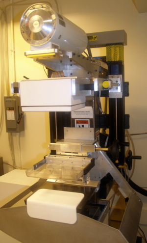
The hybrid scanner
combines two
3D modalities in the
hunt for breast cancer.
A new study suggests a hybrid scanner that unites cutting-edge 3D X-ray tomosynthesis with 3D molecular imaging tomosynthesis might help radiologists better detect hard-to-find breast cancers.
In the April issue of Radiology, scientists from the University of Virginia discussed a pilot study using their new in-house scanner. Dubbed the dual modality tomographic breast scanner (DMT), it marries three-dimensional structural and functional imaging to get results possibly several times more accurate than traditional mammography.
In X-ray tomosynthesis, a detector travels in an arc around the breast taking a series of X-ray snapshots to then render a three-dimensional image of the breast. X-ray tomosynthesis devices are currently only commercially available in Europe, but they have been generating buzz in the United States, where they might start appearing as early as next year.


Ad Statistics
Times Displayed: 10097
Times Visited: 31 Brand-New FDA-cleared Advanced Ultrasound Medical Device available for sale or lease to Wound Care Centers or any other Medical Facilities.The Arobella 1000D is designed for non-contact or debridement ultrasound wound healing therapy, or any other wounds
"Compared with 2D mammography, X-ray tomosynthesis will likely be an improvement, but it will still supply only anatomical information," says Dr. Mark B. Williams, an associate professor of radiology, biomedical engineering and physics at the University of Virginia, and lead author of the study. "We are testing the value of ganging up anatomical and functional tomosynthesis information together."
That's why in his device, Williams teams up X-ray tomosynthesis with a new technique, molecular breast imaging tomosynthesis. In this modality, a radioactive tracer, called technetium-99m sestamibi, is first injected into the patient. Cancerous tissue eats up this substance much more than healthy tissue does, so it pools together in malignancies, where it fires off gamma rays. A small gamma camera, which captures those rays, is then moved in an arc around the breast to create a series of images. Much like in X-ray tomosynthesis, the images are combined to produce a 3D molecular picture.
While each modality is effective alone, by uniting them, the DMT scanner can line up, or co-register, the structural X-ray image with the functional molecular one. The goal is to "permit the radiologist to easily identify regions of the breast in which tracer accumulation is high and know exactly where those regions are in the X-ray tomosynthesis image," Williams says.
The pilot study
To test the accuracy of the device, last year Williams and his colleagues used the DMT to scan 17 women already scheduled for breast biopsies. They then compared the results of the scans with the results of the biopsy, the so-called gold standard for diagnosing breast cancer.


