
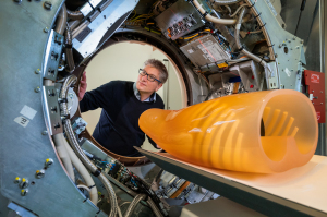
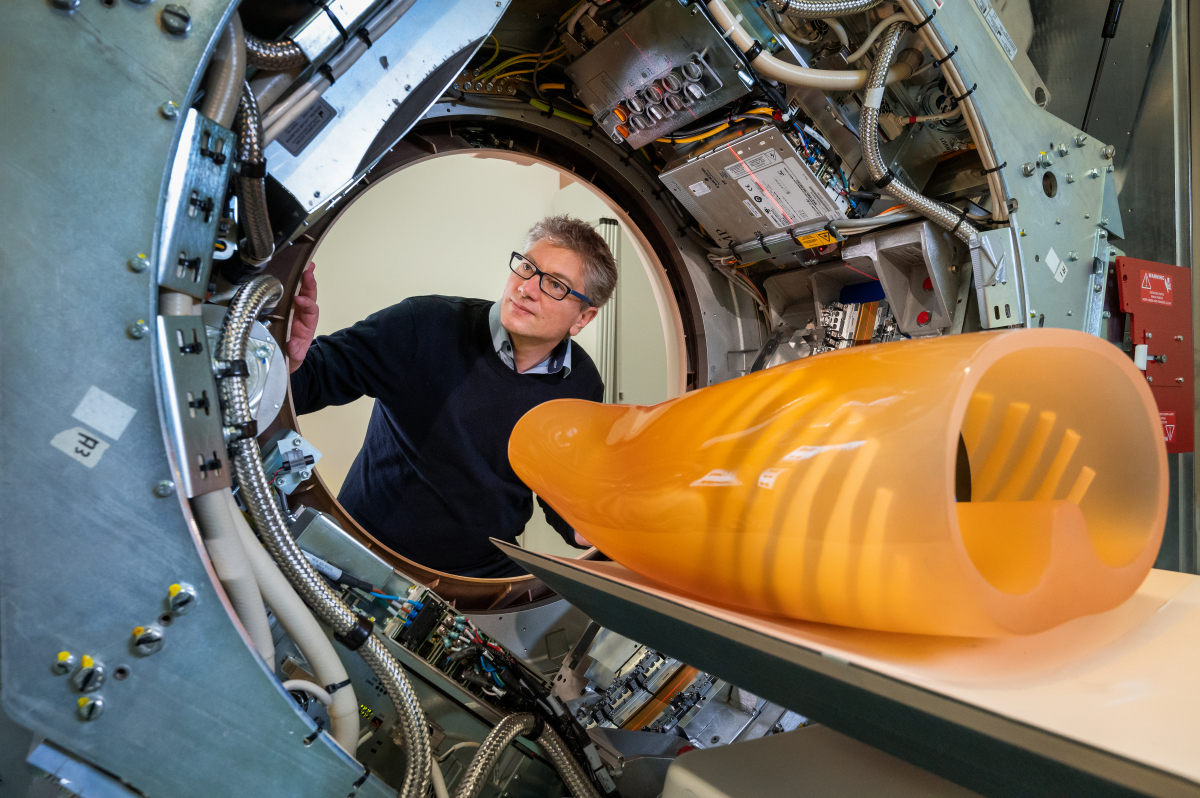
TUM researchers have developed a dark-field CT prototype to better assess human tissues (Photo courtesy of Astrid Eckert / TUM)
Researchers in Germany develop dark-field CT prototype
February 14, 2022
by John R. Fischer, Senior Reporter
Researchers at the Technical University of Munich have been able to derive more insights about human tissue for diagnosis through a combination of CT scanning and dark-field X-ray imaging.
Dark-field imaging provides information that conventional X-rays cannot on fine tissue structures, especially the lung. In conventional X-ray imaging, X-rays are attenuated by intervening tissue as they travel to the source of the detector. This creates images with varying degrees of attenuation that are based on tissue type and structures. When the X-rays interact with materials of different densities, such as an interface, they scatter. Dark-field imaging assesses this scattering effect to obtain more information on very fine tissue structures.
Technical challenges have made it hard to develop a dark-field CT device to the scale needed to assess human beings. But through their work, TUM researchers have developed a prototype that combines both technologies to produce 3D dark-field X-ray images. And it has already been used with a thorax phantom that depicts the upper human body and is large enough to repeat intended applications on real patients.
With the technique, providers can capture both conventional and dark-field X-ray images in a single scan and diagnose not just lung diseases but distinguish between various types of kidney stones and tissue deposits, authors Nikolai Gustschin and Manuel Viermetz told HCB News. "A sensitive assessment of microstructural, pathophysiological changes in tissue and their anatomical distribution provides complementary diagnostic information, which is not accessible with attenuation imaging only. It could hence facilitate a more accurate, quantitative detection and effective treatment of diseases in their early stages."
The technique relies on three optical gratings placed between the X-ray source and detector. When X-rays pass through the gratings, a characteristic pattern is produced at the detector and then changes when a person is positioned in the path of the beam. It is from these deviations that dark-field imaging is able to assess the structure of the person’s tissue. It also requires only 1/50th of the radiation dose typically used in CT scanning.
In addition to size, the fast rotation of scanning had created additional difficulties for integrating dark-field imaging. The high-speed rotation creates vibrations that affect the finely tuned components in the interior of the device. The team at TUM used them to implement the required shift between the gratings to perform dark-field imaging and developed algorithms to filter out the vibration effects based on reference scans.
The university has been assessing the approach for some time in the diagnosis of pulmonary ailments. Through research, it found that the technique can show early changes in the alveolar structure caused by chronic obstructive pulmonary disease.
"We expect the major benefit in the early diagnosis and treatment of respiratory diseases like COPD, pulmonary fibrosis, pneumonia, or lung cancer, since they are generally associated with structural changes of lung parenchyma," said Gustschin and Viermetz. "In a broader context, quantitative information on the alveolar microstructure could enable a better understanding of inflammatory processes and complications caused by acute lung injuries, infectious diseases like COVID-19 or consequences of radiation therapy."
Other potential applications, they add, include improved foreign body detection and the characterization of calcifications and bone microstructure in the context of osteoporosis.
Dark-field imaging provides information that conventional X-rays cannot on fine tissue structures, especially the lung. In conventional X-ray imaging, X-rays are attenuated by intervening tissue as they travel to the source of the detector. This creates images with varying degrees of attenuation that are based on tissue type and structures. When the X-rays interact with materials of different densities, such as an interface, they scatter. Dark-field imaging assesses this scattering effect to obtain more information on very fine tissue structures.
Technical challenges have made it hard to develop a dark-field CT device to the scale needed to assess human beings. But through their work, TUM researchers have developed a prototype that combines both technologies to produce 3D dark-field X-ray images. And it has already been used with a thorax phantom that depicts the upper human body and is large enough to repeat intended applications on real patients.
Dark-field X-ray imaging can provide more information about fine tissue structures, especially the lung
With the technique, providers can capture both conventional and dark-field X-ray images in a single scan and diagnose not just lung diseases but distinguish between various types of kidney stones and tissue deposits, authors Nikolai Gustschin and Manuel Viermetz told HCB News. "A sensitive assessment of microstructural, pathophysiological changes in tissue and their anatomical distribution provides complementary diagnostic information, which is not accessible with attenuation imaging only. It could hence facilitate a more accurate, quantitative detection and effective treatment of diseases in their early stages."
The technique relies on three optical gratings placed between the X-ray source and detector. When X-rays pass through the gratings, a characteristic pattern is produced at the detector and then changes when a person is positioned in the path of the beam. It is from these deviations that dark-field imaging is able to assess the structure of the person’s tissue. It also requires only 1/50th of the radiation dose typically used in CT scanning.
In addition to size, the fast rotation of scanning had created additional difficulties for integrating dark-field imaging. The high-speed rotation creates vibrations that affect the finely tuned components in the interior of the device. The team at TUM used them to implement the required shift between the gratings to perform dark-field imaging and developed algorithms to filter out the vibration effects based on reference scans.
The university has been assessing the approach for some time in the diagnosis of pulmonary ailments. Through research, it found that the technique can show early changes in the alveolar structure caused by chronic obstructive pulmonary disease.
"We expect the major benefit in the early diagnosis and treatment of respiratory diseases like COPD, pulmonary fibrosis, pneumonia, or lung cancer, since they are generally associated with structural changes of lung parenchyma," said Gustschin and Viermetz. "In a broader context, quantitative information on the alveolar microstructure could enable a better understanding of inflammatory processes and complications caused by acute lung injuries, infectious diseases like COVID-19 or consequences of radiation therapy."
Other potential applications, they add, include improved foreign body detection and the characterization of calcifications and bone microstructure in the context of osteoporosis.