
U.S. and Japanese researchers have developed a new technique for cross-sectional medical imaging without using tomography
New approach forms cross-section medical images without tomography
November 12, 2021
by John R. Fischer, Senior Reporter
U.S. and Japanese researchers have developed a new technique to create the first experimental cross-sectional medical image without the use of tomography.
While useful in reconstructing images from CT and PET scans, tomography can expose patients to significant amounts of radiation and slow down the speed at which scans are produced. With the new approach, radiologists can create images more quickly, potentially in real time when performing PET scans, because there is no after-the-fact reconstruction required, said Simon Cherry, professor of biomedical engineering and radiology at the University of California, Davis.
In a paper he wrote with his colleagues on the technique, Cherry says they expect the approach to make medical scanning cheaper, easier and more accurate. It relies on new, ultrafast photon detectors developed at Hamamatsu Photonics in Japan.
“When this approach can be scaled up, there is the potential for significant improvements in image quality, because more information about each gamma ray is now available and no mathematical image reconstruction is needed to create an image,” said Cherry. “Another alternative, rather than getting better images, is to get images with reduced radiation dose, or much faster (possibly even real-time nuclear medicine imaging). The approach also could lead to more compact, lower cost imaging systems.”
In PET imaging, radioactive isotopes decay and emit positrons. When these positrons come into contact with electrons in the body, they annihilate each other and emit two annihilation photons. Being able to trace the origin and trajectory of these photons theoretically creates an image of tissues. But detectors are too slow to track the photons, which prevents PET scans from capturing the full amount of data when it’s immediately available.
The researchers designed the new detectors to detect these photons (called Cherenkov photons) with an average timing precision of 32 picoseconds. This allows them to determine where the photons were created with a spatial precision of 4.8 millimeters. This, according to Cherry, is “literally imaging at the speed of light,” which allows users to produce cross-sectional images without tomography.
The detectors are composed of a Cerenkov radiator, which efficiently absorbs gamma rays and very quickly emits a small number of photons of visible light. An attached microchannel plate photomultiplier tube (MCP-PMT) converts the photons into electrons and increases the number of electrons into a detectable signal that providers can measure. The signal has a timing precision of about 25 picoseconds. Combining the MCP-PMT and Cerenkov radiator allows users to calculate the arrival time of the gamma rays within about 30 nanoseconds, according to Cherry.
Sun II Kwon, a project scientist in the UC Davis department of biomedical engineering, and Ryosuke Ota at Hamamatsu Photonics, performed experimental work with the technique in a variety of tests, including one that used an object to mimic the human brain. Cherry and his colleagues believe that with further development, the approach can be scaled to a level where it can be applied in clinical diagnostics to potentially create higher-quality images at lower radiation doses.
“The ultimate goal would be to have a scanner with the uEXPLORER geometry, but with these ultra-fast detectors,” said Cherry. “That would be an amazing diagnostic imaging device. Just as uEXPLORER was a step change over conventional scanners, this would be another step change over uEXPLORER. But, there is a long way to go before we can scale up from the two detectors in our paper, to the 500,000+ detectors in uEXPLORER.”
He adds that there will be several key developments and innovations including miniaturizing components, developing multichannel devices, developing electronics that can operate at this speed and be scalable and for the development to be cost-effective to use in a diagnostic device. Significant technology improvements are needed before the approach can be used on humans. The researchers hope to begin applying it in small animal imaging in the next two to three years.
Other collaborators included research groups led by professor Yoichi Tamagawa at the University of Fukui, and by professor Tomoyuki Hasegawa at Kitasato University.
The findings were published in Nature Photonics.
While useful in reconstructing images from CT and PET scans, tomography can expose patients to significant amounts of radiation and slow down the speed at which scans are produced. With the new approach, radiologists can create images more quickly, potentially in real time when performing PET scans, because there is no after-the-fact reconstruction required, said Simon Cherry, professor of biomedical engineering and radiology at the University of California, Davis.
In a paper he wrote with his colleagues on the technique, Cherry says they expect the approach to make medical scanning cheaper, easier and more accurate. It relies on new, ultrafast photon detectors developed at Hamamatsu Photonics in Japan.
“When this approach can be scaled up, there is the potential for significant improvements in image quality, because more information about each gamma ray is now available and no mathematical image reconstruction is needed to create an image,” said Cherry. “Another alternative, rather than getting better images, is to get images with reduced radiation dose, or much faster (possibly even real-time nuclear medicine imaging). The approach also could lead to more compact, lower cost imaging systems.”

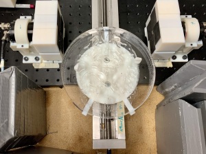
The approach was executed using new, ultrafast photon detectors
In PET imaging, radioactive isotopes decay and emit positrons. When these positrons come into contact with electrons in the body, they annihilate each other and emit two annihilation photons. Being able to trace the origin and trajectory of these photons theoretically creates an image of tissues. But detectors are too slow to track the photons, which prevents PET scans from capturing the full amount of data when it’s immediately available.
The researchers designed the new detectors to detect these photons (called Cherenkov photons) with an average timing precision of 32 picoseconds. This allows them to determine where the photons were created with a spatial precision of 4.8 millimeters. This, according to Cherry, is “literally imaging at the speed of light,” which allows users to produce cross-sectional images without tomography.
The detectors are composed of a Cerenkov radiator, which efficiently absorbs gamma rays and very quickly emits a small number of photons of visible light. An attached microchannel plate photomultiplier tube (MCP-PMT) converts the photons into electrons and increases the number of electrons into a detectable signal that providers can measure. The signal has a timing precision of about 25 picoseconds. Combining the MCP-PMT and Cerenkov radiator allows users to calculate the arrival time of the gamma rays within about 30 nanoseconds, according to Cherry.
Sun II Kwon, a project scientist in the UC Davis department of biomedical engineering, and Ryosuke Ota at Hamamatsu Photonics, performed experimental work with the technique in a variety of tests, including one that used an object to mimic the human brain. Cherry and his colleagues believe that with further development, the approach can be scaled to a level where it can be applied in clinical diagnostics to potentially create higher-quality images at lower radiation doses.
“The ultimate goal would be to have a scanner with the uEXPLORER geometry, but with these ultra-fast detectors,” said Cherry. “That would be an amazing diagnostic imaging device. Just as uEXPLORER was a step change over conventional scanners, this would be another step change over uEXPLORER. But, there is a long way to go before we can scale up from the two detectors in our paper, to the 500,000+ detectors in uEXPLORER.”
He adds that there will be several key developments and innovations including miniaturizing components, developing multichannel devices, developing electronics that can operate at this speed and be scalable and for the development to be cost-effective to use in a diagnostic device. Significant technology improvements are needed before the approach can be used on humans. The researchers hope to begin applying it in small animal imaging in the next two to three years.
Other collaborators included research groups led by professor Yoichi Tamagawa at the University of Fukui, and by professor Tomoyuki Hasegawa at Kitasato University.
The findings were published in Nature Photonics.