What's new in radiography and detectors?
November 18, 2019
by Lisa Chamoff, Contributing Reporter
Manufacturers in the radiography market have had an eye on clinician and patient satisfaction, focusing on features such as improved workflow and image quality, as well as advanced features such as digital X-ray tomosynthesis.
Meanwhile, detectors continue to get lighter and more easily maneuverable.
Here's a look at what's new.
Agfa
In February Agfa received FDA approval for the latest version of its DR 800 system.The multipurpose imager will equip providers with one solution for radiography, fluoroscopy, multi-slice and advanced clinical applications. It also extends Agfa’s capabilities from 2D single-plane imaging to include the use of tomosynthesis for synthesizing multi-slice imaging reconstruction from a single tomographic sweep.
"The DR 800 has already been supporting imaging departments to meet today's growing demand for versatility, without requiring multiple investments," said Louis Kuitenbrouwer, vice president of Agfa’s imaging division, at that time. "With the FDA 510(k) clearance for tomosynthesis, the DR 800 supports best-in-class multi-slice imaging, using algorithms that reconstruct images very quickly."
Providing iterative reconstruction, the algorithms enable faster image delivery with less noise and fewer artifacts.
Aspenstate
In January, Aspenstate began marketing its Aspen XDR straight arm digital radiography system, an entry-level X-ray room designed for the urgent care market.
When placed horizontally for a chest exam, at a 72-inch source to image distance, the straight arm measures 79 inches from end to end.
“It’s really economical solution for the urgent care market,” said Rob Scorcia, vice president of business development at Aspenstate.
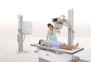
Also new to the market is the AspenFDR, a compact floor-mounted X-ray room with auto tracking for high volume patient throughput, offered with both four- and six-way tables. It’s designed for hospitals and orthopedic practices.
“It has a modern and sleek design that saves space,” Scorcia said.
Canon Medical Systems
The latest version of Canon’s Image processing software, CXDI Control Software NE, provides functionality for multi-touch monitors that enable touchscreen operations instead of a keyboard or mouse. Users can also benefit from the ID card login capability that uses near field communication technology.
Single-click Multiple Image Processing and Advanced Edge Enhancement functions for viewing of PICC lines and catheters are also part of the most recent feature release
"One-click multi-image processing supports an efficient workflow, especially in busy imaging environments," Tsuneo Imai, vice president and general manager of healthcare solutions in the business imaging solutions group for Canon USA and president of Virtual Imaging Inc.
The Advanced Edge Enhancement feature of the software is designed for evaluating soft tissue, foreign body visualization and bony detail. In addition, free rotation of images to any arbitrary angle permits fine adjustment of horizontal or vertical image orientation.
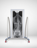
The Single Shot Long-Length Imaging feature of CXDI Control Software NE stitches long length images into a single image using multiple compatible CXDI detectors, a single x-ray exposure and the automatic stitching capability of the CXDI Software.
This new software feature, when used with compatible Canon CXDI-710C wireless and CXDI-410C wireless detectors and a specially configured stand, offers a long-length imaging workflow for whole spine or long leg exams without the need for multiple X-ray exposures. As a cost-effective solution, the detectors can be removed from the stand and used with other compatible systems
The system comes with what Imai says is a faster workflow, allowing facilities to perform more exams.
Carestream
Carestream released the new wireless DRX Plus 2530C Detector, which the company says is lighter, faster and has enhanced features.
“The newly designed small format allows for easier positioning in incubator trays,” said Jill Hamman, worldwide marketing manager for global X-ray solutions at Carestream.
The detector has an IP rating of 57, new electronic components and 98-micron pixel pitch to provide better image quality. It also features the X-Factor which enables sharing the detector across the entire DRX family of products.
At RSNA, the company will showcase the recently FDA-approved Carestream Focus 35C Detector that works with Image Suite for a wireless DR retrofit solution.
“It allows a customer to upgrade their current technology if they’re using film or CR,” Hamman said. “Not only do they get the wireless detector technology and all of the workflow improvements, but they can also get a mini PACS option. It’s a complete scheduling, image capture, archive and reporting solution.”
The solution is targeted to smaller facilities and specialty practices that are looking for an affordable way to move to wireless DR technology
The company is also rolling out its new Image View software platform, which offers a single-screen workflow for improved productivity and will run on Windows 10, allowing for additional security improvements.
“The Image View platform has an updated architecture and will allow for the support of advanced applications,” Hamman said.
Del Medical
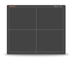
Del Medical recently launched a new detector, DELWORKS LLI, that the company says is the first full monolithic flat panel detector available in the world.
“It requires no stitching software to create a full-length image,” said Mandy Gutierrez, product marketing manager for UMG/Del Medical. “You can do a complete spine or hip to ankle in one exposure and the image displays in nine seconds, which is twice as fast as the multi-detector configurations. It eliminates issues that exist with stitching methods, like image misalignment or motion artifacts”
The 17-inch-by-42-inch detector is also more portable, wired or wireless, and can be used upright or supine, or in the OR.
“It’s for any imaging department that wants to lower X-ray dose, increase throughput and reduce exam time, with one exposure, versus up to five with other methods,” Gutierrez said.
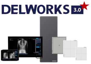
In August, the company released its DELWORKS 3.0 software, which includes the Windows 10 operating system, with enhanced security and data encryption, improved image processing and performance and the addition of enhanced image processing for lines and tubes. The software is compatible with five different detectors, including the new DELWORKS LLI 17-inch by-42-inch detector.
Fujifilm
In October, Fujifilm applied for FDA clearance for the FDR D-EVO III, what it says is the world’s first glass-free DR detector.
The detector uses film layers as its circuitry layer.
“Instead of the capture layer being adhered to glass, it’s being adhered to film technology,” said Rob Fabrizio, director of strategic marketing, digital radiography and women’s health at FUJIFILM Medical Systems U.S.A. Inc. “Glass is what makes detectors fragile and more bulky because we have to reinforce the detector … We’re making the detector a lot less fragile and we’re not sacrificing image quality.”
At less than 4 pounds, the detector is 40 percent lighter than the previous generation, D-EVO II, and the lightest in the world, Fabrizio said. It’s for portable or in-room use.
“The most advantage is going to come from portable use where it’s getting banged around and put directly under the patient,” Fabrizio said.
In mid- to late-2020, the company is also planning to release the CALNEO Dual detector, a dual capture layer detector in a portable 17-inch-by-17-inch design.
“It allows us to capture a single patient image at different layers,” Fabrizio said. “We’re able to separate out bone from soft tissue and generate three different images. In a standard chest X-ray, you can separate out the bone and better see a spot in the lung field that could be hidden by the ribs. If it disappears in soft tissue image it’s likely calcified, and not cancerous. For patients with a known spot on their lung, this can also be an easier way to track it. It’s significantly faster, lower dose and lower cost than traditional imaging, such as CT, for that type of exam.”
Fujifilm is also releasing three new radiography rooms, two of which will be showcased at RSNA.
The FDR Clinica-U is a compact single detector U-arm system for orthopedic practices and where space and power requirements are limited.
“This is a simple, cost effective way to install DR with a single detector positioning system,” Fabrizio said.
The tube stays directly aligned with the detector at all times and the system bolts to floor, so it doesn’t require ceiling prep.
The FDR Clinica-X is a mid-range overhead system for general radiography and ER. It has an overhead tube system, chest stand and table. The room will have the ability for stitching and auto positioning with later releases.
“It’s a general grab and go type of room,” Fabrizio said. “There’s a lot more focus toward having advanced features in the mid-range and on the low-end rooms. These new rooms are targeted as more cost-effective solutions without sacrificing the latest in functionality or performance.”
Clinica-U and Clinica-X are not FDA cleared.
The FDA-cleared D-EVO Suite FSX is a floor-mounted room designed for the hospital and outpatient environment. It has auto tracking, advanced tube head display and other premium features normally associated with an overhead room.
“Hospitals use them in rooms that are too small for an overhead room,” Fabrizio said.
GE Healthcare
GE Healthcare’s newest radiography release is its FlashPad HD, for use with the Discovery XR656 HD and Optima XR646 HD X-ray systems, and Optima XR240amx mobile X-ray system.
The 17-inch-by-17-inch detector comes in the full bucky size, so there is no need to rotate for portrait or landscape aspects.
“That really satisfies a large install base of folks who wanted that large square detector,” said Todd Minnigh, chief marketing officer for X-ray for GE Healthcare.
GE has advanced applications, such as VolumeRAD and Dual Energy, that are now available for these HD detector-based systems.
In September, the FDA cleared GE’s Critical Care Suite, which has an algorithm that flags a likely pneumothorax finding as well as AI algorithms to automatically reorient the image, identify incorrect protocol selection and highlight potentially clipped anatomy on chest images.
Konica Minolta
Konica Minolta has developed a technology called Dynamic Digital Radiography, a burst of continuous X-ray exposures that acquires 15 images per second for up to 20 seconds, similar to fluoroscopy but not in real time.
The company has partnered with Shimadzu to bring Dynamic Digital Radiography to the market on the RADSpeed Pro radiographic imaging system.
“It’s better resolution than fluoro for bones and soft tissue without contrast,” said Guillermo Sander, director of marketing and digital radiography for Konica Minolta Healthcare Americas. “And because its digital radiography, we can put metrics on it. We can calculate the lung volume and provide numbers and graphs to help with the diagnosis. We believe that by using DDR instead of static chest X-rays, clinicians will be able to rule out more things in less time. We really think it’s helping bring more information to a modality that has been very stable for a long time.”
Philips
At last year’s RSNA, Philips released the DigitalDiagnost C90, a ceiling-suspended radiography system available in multiple configurations and featuring a camera with a birds-eye view of the patient.
Stefan Mintert, senior portfolio manager for DXR at Philips, said it’s the first system on the market with a live camera and touchscreen monitor at the tube head. It is designed to avoid retakes as the staff can check the patient is correctly positioned from the tableside.
“It solves issues from all our stakeholders,” Mintert said. “In radiography, efficiency is the main driver because it’s about patient throughput and exams done as quickly as possible.”
Rayence
In 2020, Rayence is going to be introducing a dynamic detector that’s a replacement for an image intensifier in radiography and fluoroscopy rooms.
The panel uses indium gallium zinc oxide (IGZO) and there will be cesium iodine in the receptor.
It will be shown at this year’s RSNA.
“The detector has technology that helps with the speed and quality of the image,” said Ed Terzi, marketing manager for Rayence USA. “With fluoroscopy, you're looking at higher frame rates.”
The dynamic panels come in 6-, 9-, 12-, 16- or 17-inch sizes.
The company has also made improvements to its current thin-film transistor TFT panels to reduce radiation dose.
Rayence also released a new version of its image processing software, Xmaru Pro, which provides further processing of the image for specific applications.
“It allows for more understanding of what's going on in certain areas, like the lung,” Terzi said.
Samsung
In July, Samsung launched two new products as part of a new platform called iQuia. It includes new 14x17 and 17x17 wireless detectors for fixed and mobile systems. The exterior housing of the detectors was redesigned to give it a more ergonomic feel with tapered edges and the distributed weight capacity was increased to 882 pounds.
As part of the launch there is also an enterprise management tool called SMART Center by iQuia.
“SMART Center allows us to aggregate multiple different metrics from the site’s install base so radiology directors, QA/QC techs, and clinical engineers are able to manage their systems,” said Boris Geyzer, product manager for digital radiography at Samsung.
Additionally, SMART Center manages dose metrics from the Samsung systems to help facilitate staff training for studies that have higher deviation index numbers.
The company has also updated its floor-mounted system from the GF50 to GF55 for the value and performance markets.
Lastly, Samsung just finished the launch of new co-marketing effort with Del Medical, marrying Del’s ceiling and floor mount radiographic systems with Samsung’s detectors and image acquisition software, providing Samsung customers a full series of products from the fully robotic GC85A ceiling system to a manual ceiling system from Del.
All products are compatible with Samsung’s full suite of software, including Bone Suppression, a deep learning AI algorithm that eliminates the ribs and clavicle from a chest image for better visualization of lung nodules.
Shimadzu
In 2019, Shimadzu released three new products.
The SONIALVISION G4 LX edition is the newest version of the company’s universal remote RF table, offering an optional tube SID that extends up to 180 centimeters, and several other features.
The 180 cm SID is geared toward both price-sensitive buyers and/or space-limited X-ray rooms, said Frank Serrao, marketing manager for Shimadzu.
“Institutions with a tight budget can save money by not ordering an overhead X-ray tube system but can still maintain standard 180cm exposures using the LX,” Serrao said. “The same would apply to institutions that just don’t have the space for an overhead tube system.”
In August, the FDA cleared the FLUOROspeed X1, a traditional RF table with patient-side controls. In traditional RF systems used in the U.S., the X-ray tube is mounted below the table and the detector sits on a deck above the patient.
“The operator does all the X-ray acquisitions right from the table deck controls,” Serrao said. “The deck moves fluidly with an ambidextrous control handle which has both Rad and RF exposure buttons available at the fingertips.”
The X1 elevating table can be lowered to the ground for easier access while accommodating patients with large girths having 31.5 inches of aperture between the tabletop and the deck, which Serrao believes is the largest in the industry.
Additionally, a new Konica Minolta technology called Dynamic Digital Radiography, or DDR, is an advanced application for use on the RADspeed Pro radiography room system and is ideal for imaging pneumothorax and various chest functions.
“DDR is a fast first-line test that combines anatomic and functional information generated by the rapid acquisition of a series of radiographs reconstructed from algorithms outputting a variety of metrics to help clinicians make informed decisions,” said Charles Cassudakis, rad and RF director at Shimadzu.
“Partnering with Konica Minolta to develop and distribute the DDR system proved to be the ideal scenario because of Konica Minolta’s film technology background,” Serrao said.
Siemens Healthineers
In January, the FDA cleared the Multix Impact, a floor-mounted DR system from Siemens Healthineers for cost-conscious facilities.
It features a touchscreen user interface to allow the technologist to stay with the patient for longer periods of time, with avatars and job aids to help technologists if they come across an exam they haven’t seen before.
There is also a flat, free-floating tabletop for easier patient access and tube tracking between the detector and X-ray tube.
“You typically only see this on high-end units,” says Joseph D’Antonio, senior director of product management and marketing for X-ray products and women’s health at Siemens Healthineers North America.
In March, the FDA cleared the Mobilett Elara Max, which runs on a Windows 10 platform for more hardened cybersecurity, 180-degree lateral arm movement and a virtual workstation for access to a PACS and RIS system during an exam, so the technologist can sign off on the exam in the field.
“You can see the patient record on the screen of the mobile device,” D’Antonio said.
Swissray
At this year’s RSNA, Swissray will be showing the ddRAura S, a motorized straight arm that can be used with a variety of tubes and detectors.
They will also show the ddRAura Air, which will have its U.S. debut in the first quarter of 2020. The Air is a fully automated, dual-OTC system, with the first column carrying the tube and collimator and the second hosting a dynamic panel.
“The beauty of the system is you can use it as a wall stand, perform any off-table exams, or use it with the 90/90 table as a remote-control RF system,” said Ed Pol, chief executive officer of Swissray Customer Care.
This is a premium product for hospitals, imaging centers and orthopedics.
“We call it our all-in-one system. It allows the user to do a variety of exams that would typically require the use of different rooms to perform ,” Pol said.
Swissray’s first radiography and fluoroscopy room has dual-size fluoroscopy capabilities, with a 17-inch and 11-inch mode.
“It works very much like conventional RF system, with the ease and convenience of programmable, automated, robotic positioning and the capability of acquiring full size, full resolution radiographs at speeds up to four frames per second,” Pol said.
Thales
In June, Thales released the ArtPix DRF, a software platform for radiography and fluoroscopy that provides full HD images and advanced clinical applications, including stitching, digital X-ray tomosynthesis and a color-coded exposure index map.
Customers have the option of a tablet user interface so the clinician can stay close to the patient.
The solution also has a customizable workflow that can also be built around the organization's branding.
"We think it will bring a clear differentiator to the clinician," said Camille Vigier, product line manager for X-ray products at Thales.
The official U.S. launch will be at this year's RSNA.
Varex Imaging
Earlier this year, Varex Imaging released a new version of the 4336Wv4 wireless detector, with a reduced weight, improved ergonomics, and with a new long-life battery.
The company is also working on releasing a 17-inch-by-17-inch version, the 4343W, early next year. It’s expected to weigh 3.2 kilograms and has an IP rating of IP67.
“These days, the weight and ergonomics are really important, so we improved those things,” said Tuomas Holma, product manager for X-ray detectors at Varex Imaging.

