by
John R. Fischer, Senior Reporter | February 16, 2024
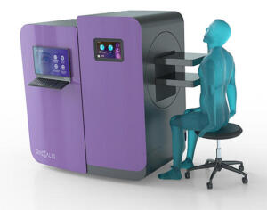
Radialis PET Imaging scanner
Among the large population of women with dense breast tissue, many find it challenging or are unable to undergo breast MR due to claustrophobia and contraindications. Now, researchers in Toronto, Canada, are saying that an alternative may be available that is equal to MR scan quality and with fewer false positives and radiation dose levels equivalent to standard mammography.
With lower false positives and strong sensitivity, low-dose positron emission mammography (PEM) has the potential to prevent unnecessary follow-ups better than MR imaging, according to the authors. It also eliminates breast compression required with traditional mammography, making it more comfortable for patients.
In a study of 25 women recently diagnosed with breast cancer, the technique identified 24 of the cases with invasive cancer (96%). Its false positive rate was 16%, compared to 62% with MR imaging. The researchers performed PEM with fluorine 18-labeled fluorodeoxyglucose (18F-FDG) and Radialis' organ-targeted PET imaging scanner. Two breast radiologists reviewed the images taken one and four hours after the 18F-FDG injection and correlated findings with lab results.


Ad Statistics
Times Displayed: 174441
Times Visited: 3180 For those who need to move fast and expand clinical capabilities -- and would love new equipment -- the uCT 550 Advance offers a new fully configured 80-slice CT in up to 2 weeks with routine maintenance and parts and Software Upgrades for Life™ included.
“The integration of these features — high sensitivity, lower false-positive rates, cost-efficiency, acceptable radiation levels without compression, and independence from breast density — positions this emerging imaging modality as a potential groundbreaking advancement in the early detection of breast cancer,” said lead author Dr. Vivianne Freitas, assistant professor at the University of Toronto, in a statement.
The Radialis PET Imager positions organs between two planar detectors, which can be rotated and reach across the body or head to scan the area of interest. The detectors can be raised or lowered to image patients sitting, standing, or lying down. It also can show objects as small as 1.3mm; use more available data to reveal clinically relevant details not visible with traditional PET; and distinguish between adjacent areas of relatively similar radiotracer uptake.
According to Freitas, the method could potentially be used for screenings and diagnostic purposes, helping to address limitations with mammography, which may be obscured from detecting tumors by dense breast tissue. It also could be used to interpret uncertain mammogram results, assess chemotherapy responses, and determine the extent of the disease in newly diagnosed breast cancer, including involvement of the other breast.
More studies are required to test PEM’s efficiency and the potential role it could play in clinical practice. Freitas is currently evaluating its ability to reduce high rates of false positives typically associated with MR scans and says that if it can do this, the solution could significantly reduce discomfort and anxiety linked to these events as well as unnecessary biopsies and treatments.
“While the full integration of this imaging method into clinical practice is yet to be confirmed, the preliminary findings of this research are promising, particularly in demonstrating the capability of detecting invasive breast cancer with low doses of fluorine-18-labeled FDG,” Dr. Freitas said.
The findings were published in
Radiology: Imaging Cancer, a journal of the Radiological Society of North America (RSNA).