by
John R. Fischer, Senior Reporter | May 18, 2020
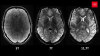
MR scans of COVID-19 patients in the ICU showed brain abnormalities, including signs of encephalitis.
A new study has found that close to half of COVID-19 patients admitted to ICUs in Turkey showed signs of brain abnormalities on MR scans.
The discovery was made by neuroradiologists at Istanbul University-Cerrahpasa, who assert that the findings validate symptoms already identified for those with the disease, particularly symptoms related to the nervous system.
“Recent evidence highlights a relatively high percentage (36%) of central nervous system symptoms including headache, altered mental status, acute cerebrovascular disease and epilepsy in patients with COVID-19,” said the authors in their report. “The rate of neurological symptoms is higher in patients with more severe respiratory disease status. The relatively high percentage of neurologic symptoms is concordant with studies showing neurotropism of coronavirus.”


Ad Statistics
Times Displayed: 173425
Times Visited: 3165 For those who need to move fast and expand clinical capabilities -- and would love new equipment -- the uCT 550 Advance offers a new fully configured 80-slice CT in up to 2 weeks with routine maintenance and parts and Software Upgrades for Life™ included.
The purpose of the study was to evaluate the neurological condition of COVID-19 patients, as current literature on the subject is limited. Six percent of patients assessed experienced acute stroke, while another 15% had an altered state of mind. The authors believe one factor responsible is the cytokine storm, in which the body attacks its own cells and tissues in addition to fighting off the virus,
according to WebMD.
The researchers performed MR exams on 235 COVID-19 patients in the ICUs of two academic and six non-academic-affiliated hospitals. Of them, 50 (21%) developed neurological symptoms, with 27 examined under brain MR. Of these 27, 12 (44%) showed acute findings. Ten had COVID-19-related neuroimaging findings that showed cortical FLAIR signal abnormalities, including an increased cortical diffusion-weighted signal with corresponding low ADC values, subtle leptomeningeal development and punctate cortical blooming artifact.
All abnormalities were found in different areas of the brain, including the frontal lobe, parietal lobe, occipital lobe, temporal lobe, insular cortex and cingulate gyrus. Diagnoses that included autoimmune encephalitis, seizure, hypoglycemia and hypoxia were the main differentiator of the group, as it can be accompanied by cortical microhemorrhages and blood-brain barrier breakdown. The findings highlight that COVID-19 may result in infectious or autoimmune encephalitis due to its neurotropic potential.
"Certain viral and autoimmune encephalitis can have a specific pattern of involvement that is helpful to establish a differential list,” wrote the researchers. “However, non-specific imaging patterns in our series hinders achieving a specific diagnosis based on MR.”