by
John R. Fischer, Senior Reporter | December 18, 2018
Using ghost imaging could reduce
exposure to radiation during 3D X-ray
exams
A collaboration spanning both hemispheres has uncovered a new way to reduce the amount of exposure that patients are subjected to under 3D X-ray medical scanning.
Working alongside the European Synchrotron Radiation Facility, researchers at Australian National University and Monash University found that using a technique called ghost imaging could enable scientists to retrieve 3D X-ray images of the interior of objects that are opaque to visible light, with only the total X-ray transmission measured by a single sensor. The teams say that a variation of the approach could require only a sensor rather an X-ray camera.
“Because of the potential for significantly lower doses of X-rays with 3D ghost imaging, this approach could revolutionize medical imaging by making X-ray screening for early signs of disease much cheaper, more readily available and able to be undertaken much more often,” Dr. David Paganin, a physicist at Monash University in Australia and the senior author of the research paper behind the finding, said in a statement. “This would greatly improve early detection of diseases, including cancers.”


Ad Statistics
Times Displayed: 172546
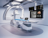
Times Visited: 3115 For those who need to move fast and expand clinical capabilities -- and would love new equipment -- the uCT 550 Advance offers a new fully configured 80-slice CT in up to 2 weeks with routine maintenance and parts and Software Upgrades for Life™ included.
Current 3D X-ray approaches are limited due to increased risks for cancer from prolonged exposures to damaging X-rays. This prevents patients from undergoing CT and mammography procedures as frequently as they may need to, for serious conditions such as breast cancer.
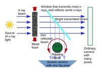
The application of ghost imaging correlates two beams that do not individually carry any meaningful information about an object. One beam encodes a random pattern that acts as a reference and never directly probes the sample, while the other does pass through it.
The researchers tested their hypothesis by creating X-ray patterns that shone a bright beam of X-ray light through a metal foam. They then took a 2D image of the random beam and passed a very weak copy of it through the sample. A large-area, single-pixel detector captured the X-rays that passed through it, with the process repeated for multiple illuminating patterns and sample-object orientations to construct a 3D tomographic image of the internal structure of the object.
The teams then tested the approach in a proof-of-concept experiment involving an aluminum cylinder with a diameter of 5.6 millimeters and two holes of less than two-millimeter diameter. The team produced 3D images with 1.4 million voxels that carried a resolution of 48 millionths of a meter.
“X-ray ghost imaging, especially ghost tomography, is a completely new field that needs to be explored and developed much further,” Dr. Andrew Kingston, a postdoctoral fellow at the Australian National University, said in a statement. “With more development, we envision ghost X-ray tomography as a route to cheaper and, therefore, much more readily available 3D X-ray imaging machines for medical imaging, industrial imaging, security screening and surveillance.”
Numerous efforts to enhance the use and quality of X-ray imaging have been underway this year, including a recent example in which a father-son duo from New Zealand
performed the first 3D color X-ray using CERN technology.
The paper was published in the journal,
Optica.