New approach promises rapid 3D model production
by
John R. Fischer, Senior Reporter | July 09, 2018
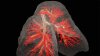
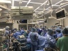
A new approach may provide quicker
3D production with greater accuracy
in anatomical features
Clinicians utilizing individual-based 3D printed models may soon find greater accuracy in their anatomical compositions.
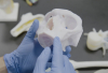
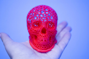
A team of international researchers has developed a new approach that leverages the potential of patient-specific medical data to rapidly produce more realistic detailed physical models, in the process saving on time required with conventional 3D printing and instilling greater confidence in performing surgical procedures.
“To date, virtually all 3D-printable medical data sets are created using traditional image thresholding, subsequent isosurface extraction, and the generation of .stl surface mesh file formats,” the researchers said in their study. “These existing methods, however, are highly prone to segmentation artifacts that either over- or under-exaggerate the features of interest, thus resulting in anatomically inaccurate 3D prints. In addition, they often omit finer detailed structures and require time- and labor-intensive processes to visually verify their accuracy.”
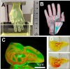
Deriving information from volumetric data sets, researchers developed a bitmap-based workflow and utilized cross-sectional stacks of images acquired for diagnostic purposes or in advance of surgical operations, applying standard photo editing workflow to each bitmap slice.
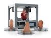
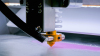
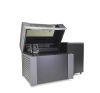
Slices were then fed directly into an inkjet-like 3-D printer to produce models incorporating grayscale, transparency and mechanical property gradients to closely mimic patient-specific anatomy.
Open-source software equipped with existing image-processing algorithms was applied in the preparation of bitmap input files, which would allow for widespread adoption of the method. In addition, modification of workflows would enable further experimentation with exploration of different window adjustments and dithering algorithms in the production of fully customizable and application-specific 3D prints.
Limitations do exist within the technology, however, characterized by inaccuracies from image reconstruction methods, the need to modify traditional clinical tomography file compression and archival protocols, data set-specific issues, and the inherent resolution of imaging modalities.
But the team says that such errors can be addressed and that, with more research, their technique may improve treatment options and lead to new areas of study in precision medicine.
“By lowering barriers to the visualization of fine details in biorealistic 3D-printed models, we hope to broaden access to this technology for a wide range of medical professionals and patients,” said the authors. “When combined with high-resolution biological imaging data, multimaterial medical 3D printing has the potential to improve treatment, enhance communication, and open new research avenues in precision medicine.”
Coauthors include James Weaver, Wyss Institute for Biologically Inspired Engineering, Harvard University (Cambridge, Massachusetts); and colleagues from MIT (Cambridge, Massachusetts); Massachusetts General Hospital and Harvard Medical School (Boston, Massachusetts); University of Washington (Seattle, Washington); Brigham and Women's Hospital (Boston, Massachusetts); Isomics (Cambridge, Massachusetts); Universitätsmedizin Berlin (Berlin, Germany); Beth Israel Deaconess Medical Center (Boston, Massachusetts); and Max Planck Institute of Colloids and Interfaces (Potsdam, Germany).
The findings were published in 3D Printing and Additive Manufacturing, a peer-reviewed journal from Mary Ann Lieber, Inc., publishers.
Researchers and Mary Ann Liebert, Inc., publishers did not respond for comment.
|
|
|
You Must Be Logged In To Post A Comment
|