At South Denver Neurosurgery,
intraoperative technology means doing
in two hours what once took 10
From the April 2018 issue of HealthCare Business News magazine
By Dr. David VanSickle
Deep brain stimulation, also known as DBS, is a surgical procedure that is far from new.
It’s been around for nearly 40 years. However, the technique has remained primarily the same for much of its existence.


Ad Statistics
Times Displayed: 174790
Times Visited: 3187 For those who need to move fast and expand clinical capabilities -- and would love new equipment -- the uCT 550 Advance offers a new fully configured 80-slice CT in up to 2 weeks with routine maintenance and parts and Software Upgrades for Life™ included.
As technology continues to evolve, it is crucial that we, as surgeons, continue to enhance our techniques in order to make surgeries such as DBS more efficient, affordable and accessible to those who need it most. Our current health care environment demands it.
An evolving procedure
DBS is a neurological procedure primarily used to treat those with Parkinson’s disease. The surgery has the ability to rewind symptoms roughly seven years by reducing the motor symptoms of Parkinson’s such as slowness, tremors and shuffling, while simultaneously improving facial expression, rigidity and fine and coarse motor skills. On average, only 2 to 10 percent of those who need DBS ever have the opportunity to receive it. One of the major reasons for that is if a hospital is outside of an academic institution, it is simply unaffordable for both the hospital and the patient. With today’s health care system, the reimbursement rates from insurance companies are lower, limiting the ability for hospitals to provide care.
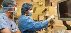
For more than 30 years, surgeons performed DBS the same way. The process, known as microelectrode mapping, requires the surgeon to place a headframe, acquire MR and CT images and then line up to the target. From there, the surgeon creates a burr hole and passes fine wire microelectrodes through the brain multiple times, recording cell firings to create a map of the brain from a physiologic point of view.
The surgeon then places the lead and, while the patient is awake, fine-tunes the location, and passes an electrode down to test it. While testing the electrode, the surgeon uses physiologic majors to improve the symptoms. Once dialed in, the electrode stays in place permanently. The process can take eight to 10 hours and is typically done over the course of two days, and sometimes separated by months or even years.
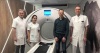
In 2010, Dr. Kim Burchiel of the Oregon Health & Science University wanted to develop a new technique that would allow surgeons to perform DBS under anesthesia. In order to do this, he needed to find the leads in their proper locations, which he solved by using a CT system. Dr. Burchiel was the first surgeon to use the CereTom, a portable CT system, in an operating room in order to place a lead. He realized that this technique, which focused on anatomy rather than physiology, worked just as well. He used live images fused with a preoperative MR in order to check where the lead was. By following this technique, he was able to eliminate the physiological testing completely and rely on the accuracy of the CereTom images.