by
Lauren Dubinsky, Senior Reporter | August 22, 2017
May also monitor effectiveness
of drugs
A new diagnostic tool that uses near-infrared light may be able to detect a patient’s risk of heart attack and stroke.
The tool, which was developed by WMG, University of Warwick, the Baker Institute and Monash University, identifies high-risk arterial plaques.
"The discovery of this autofluorescence of the unstable atherosclerotic plaques is very characteristic for these dangerous plaques," Dr. Tara Schiller of WMG, University of Warwick and Dr. Karlheinz Peter of the Baker Institute, wrote to HCB News in an email. "The signal is only seen in these plaques."
The research team found that when they increased the wavelength of the light currently used to assess fatty build-up in arteries, they could selectively spot the rupture-prone deposits that typically lead to blood clots, heart attacks and strokes.
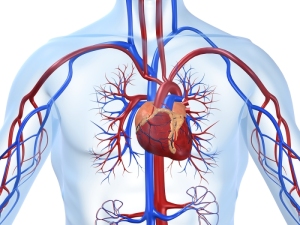
Cardiovascular disease accounted for almost 801,000 deaths in the U.S. this year, according to the American Heart Association. These diseases claim more lives each year than all types of cancer and chronic lower respiratory disease combined.
Peter stated that when they shine a light in the near-infrared wavelength range, the light is reflected at a certain wavelength. So essentially, they can use laser light to illuminate the plaques that are unstable.
The products that caused this fluorescence were identified using Raman spectroscopy. They’re thought to be a mixture of heme products that form when red blood cells degrade.
These products were only observed in unstable plaques with internal bleeding, and not in the more stable fatty deposits. That helps to improve selectivity when searching for high-risk deposits and may help physicians spot the highest-risk patients.
Current imaging techniques are capable of identifying some characteristics of high-risk plaques, but none have been broadly recognized as reliable methods for selectively detecting these types of plaques.
"Coronary angiography can only detect narrow segments in the coronary artery, but it does not report on inflamed and thereby rupture-prone plaques that sit in the wall of the coronary arteries," said Peter.
This new imaging technique needs to be tested in clinical trials, but if the results are promising, it could be used to evaluate unstable fatty arterial plaques. It also has the potential to monitor the effectiveness of drugs intended to prevent heart attack and stroke.
Schiller added that they need to develop a technology that allows them to evaluate the coronary arteries of patients. The current standard technique uses plaques removed from a patient's carotid artery, and all patients are anonymous but alive after surgery.
Back to HCB News