Scientists create MR technology that improves image quality and reduces acquisition time
by Lauren Dubinsky, Senior Reporter | May 30, 2017
MRI

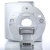
A simulation of a patient undergoing
a MR scan modified by metasurface
a MR scan modified by metasurface
Scientists from the Netherlands and Russia developed a metasurface-based technology for MR scanners that has the potential to either reduce acquisition time or improve image quality.
MR imaging provides valuable structural and functional information, but it requires more time than CT or ultrasound to acquire an image because of its low signal-to-noise ratio.
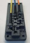
The metasurface, which is composed of conducting copper strips, is positioned between the patient and the receive coil arrays during the exam to enhance the signal-to-noise ratio in the region of interest.


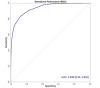
 The scientists from Leiden University Medical Center and ITMO University tested the metasurface on human patients and found that it increases local sensitivity by 50 percent. The results of the study were published in Scientific Reports.
MR scans often have to be repeated many times, and the signals then need to be added together. However, the scientists found that the metasurface obviates that.
"Conventionally, if an examination now takes twenty minutes, it may only need ten in the future," Alexey Slobozhanyuk, research fellow at ITMO University, said in a statement. "If today hospitals serve ten patients a day, they will be able to serve twenty with our development."
Alternatively, instead of accelerating the procedure, the metasurface could be used to increase the image resolution, since the size of the 3-D pixel is also limited by the signal-to-noise ratio.
Historically, it has been a challenge to create a technology like this because the dimensions of the metamaterials were too large to integrate into close-fitting receive coil arrays. The scientists solved that by pioneering an ultra-thin design.
"Our technology can be applied for producing metamaterial-inspired ultra-thin devices for many different types of MRI scans, but in each case, one should, first, carry out a series of computer simulations as we have done in this work," said Rita Schmidt, first author of the study and researcher at Leiden University Medical Center. "One needs to make sure that the metasurface is appropriately coupled."
MR imaging provides valuable structural and functional information, but it requires more time than CT or ultrasound to acquire an image because of its low signal-to-noise ratio.
The metasurface, which is composed of conducting copper strips, is positioned between the patient and the receive coil arrays during the exam to enhance the signal-to-noise ratio in the region of interest.

New Fully Configured 80-slice CT in 2 weeks with Software Upgrades for Life
For those who need to move fast and expand clinical capabilities -- and would love new equipment -- the uCT 550 Advance offers a new fully configured 80-slice CT in up to 2 weeks with routine maintenance and parts and Software Upgrades for Life™ included.
MR scans often have to be repeated many times, and the signals then need to be added together. However, the scientists found that the metasurface obviates that.
"Conventionally, if an examination now takes twenty minutes, it may only need ten in the future," Alexey Slobozhanyuk, research fellow at ITMO University, said in a statement. "If today hospitals serve ten patients a day, they will be able to serve twenty with our development."
Alternatively, instead of accelerating the procedure, the metasurface could be used to increase the image resolution, since the size of the 3-D pixel is also limited by the signal-to-noise ratio.
Historically, it has been a challenge to create a technology like this because the dimensions of the metamaterials were too large to integrate into close-fitting receive coil arrays. The scientists solved that by pioneering an ultra-thin design.
"Our technology can be applied for producing metamaterial-inspired ultra-thin devices for many different types of MRI scans, but in each case, one should, first, carry out a series of computer simulations as we have done in this work," said Rita Schmidt, first author of the study and researcher at Leiden University Medical Center. "One needs to make sure that the metasurface is appropriately coupled."
You Must Be Logged In To Post A CommentRegisterRegistration is Free and Easy. Enjoy the benefits of The World's Leading New & Used Medical Equipment Marketplace. Register Now! |
|