by
Lisa Chamoff, Contributing Reporter | May 01, 2017
MR imaging, 3-D printing and a bit of Hollywood magic helped a team of neurosurgeons, computer engineers and special effects experts create a way for surgeons to practice minimally invasive brain surgery.
The effort was recounted in an article recently published in the
Journal of Neurosurgery: Pediatrics.
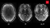
After an MR scan, the team created a full-scale reproduction of the head of a 14-year-old patient with hydrocephalus, a buildup of cerebrospinal fluid in the brain. After the brain, scull and scalp were 3-D printed, the special effects team brought in softer materials that mimicked the texture of the external and internal tissue, and the model included a basilar artery and ventricles that pulsated, and cerebrospinal fluid that moved in a realistic way.


Ad Statistics
Times Displayed: 174517
Times Visited: 3183 For those who need to move fast and expand clinical capabilities -- and would love new equipment -- the uCT 550 Advance offers a new fully configured 80-slice CT in up to 2 weeks with routine maintenance and parts and Software Upgrades for Life™ included.
The patient looked so realistic that the journal said it needed consent to include their picture, said Dr. Alan Cohen, chief of pediatric neurology at Johns Hopkins Hospital and a senior author of the report.
A group of four neurosurgery fellows and 13 residents used the patient model to perform endoscopic third ventriculostomy (ETV), in which a small hole is created for the fluid to flow through, eliminating the need for implantation of a shunt, which comes with risks. ETV is performed via a small hole in the patient’s skill using an endoscope.
The trainees then rated the model on a five-point scale, with an average rating of 4.88 for the model’s effectiveness in training. The trainees’ performances were also rated, and the fellows received higher scores, showing that the model can distinguish between levels of surgical experience.
There have not been good ways to practice these new minimally-invasive surgical methods, Cohen said. Cadavers are expensive and not reusable, and the cause of death is usually not the problem the surgeons are looking to treat. They’ve also tried replicating surgery techniques using bell peppers and pumpkins, and used virtual reality, but none of the methods are realistic enough.
“It’s a different eye-hand coordination,” Cohen told HCB News. “We thought there was a need for a better technique.”
The model can be used for different types of brain surgery, with other components able to be added.
Cohen said that while the initial model is expensive, and a facility would need a 3-D printer to create the components, it is better than the usual teaching methods.
“I think it’s like computers,” Cohen said. “When they first come out, they’re more expensive, but the price gets lower. I think this will not only replace how we teach minimally invasive neurosurgery, but all neurosurgery.”
Back to HCB News