by
Christina Hwang, Contributing Reporter | October 19, 2016
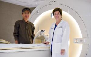
Dr. Changho Choi and Dr. Elizabeth Maher
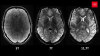
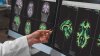
Using MR spectroscopy, researchers at UT Southwestern Medical Center have found a new biomarker, 2HG, to track certain tumors that occur in the brain.
MR spectroscopy, a non-invasive method to measure biochemical changes in the brain, compares the chemical composition of normal brain tissue with genetically mutated tissue isocitrate dehydrogenase (IDH), and also aids in measuring 2HG concentration during cancer progression.
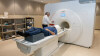
In their study, 136 patients were scanned with MR. Sixty patients came in once for a scan and the remaining were scanned multiple times and followed during the course of their disease.


Ad Statistics
Times Displayed: 174512
Times Visited: 3183 For those who need to move fast and expand clinical capabilities -- and would love new equipment -- the uCT 550 Advance offers a new fully configured 80-slice CT in up to 2 weeks with routine maintenance and parts and Software Upgrades for Life™ included.
The researchers used MR spectroscopy to detect 2HG in the patients to help them identify cancerous cells since 2HG is not elevated in normal cells.
“We can measure 2HG concentration and follow it during the disease course,” senior author Dr. Elizabeth A. Maher, associate professor of internal medicine, and neurology and neurotherapeutics, told HCB News.
“It's stable over long periods of time when the tumor is stable, increases when the disease progresses, and is an excellent marker of tumor response because it falls in response to treatment that is working,” she said.
Using the 2HG biomarker, physicians can see if the drug is able to reach the tumor and inhibit it within a week. If it inhibits the tumor, the 2HG concentration should decrease within a few days.
Before the new biomarker was used, the physicians would not know if the drug was able to inhibit the target and they could only hope and try to look for a response, Maher said.
The study builds off of previous research from UT Southwestern and the Advanced Imaging Research Center. In the 2012 study, the investigators discovered that they could detect 2HG in a tumor with high sensitivity and specificity. In this study, researchers showed that 2HG can help track the disease.
Currently, MR spectroscopy and 2HG are being used to learn more about glioma, the most common type of brain cancer. The researchers are learning more about how the tumor grows, how it responds to treatment and how it becomes resistant.
Lead author Dr. Changho Choi, professor of radiology and part of the Advanced Imaging Research Center, noted that 2HG could also be useful in diagnosing brain tumors where surgery to obtain tissue samples is not an option, such as inaccessibility of the tumor or when obtaining a sample could cause neurological damage.
“We established in this study that 2HG levels in these tumors can be used to make a 'presumptive' molecular diagnosis of an IDH mutation, based solely on imaging,” Choi said, in a statement.
Maher credited the patients for their commitment to the research.
“This is really a terrific story about how patients partner with doctors and scientists to advance science and especially translational medicine,” she said. “We will be getting this method out to any radiology department that wants to do this imaging and we have the patients to thank for making it happen.”