by
Lauren Dubinsky, Senior Reporter | April 28, 2015
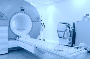
Johns Hopkins researchers have developed a new imaging technique that combines standard MRI scans with motion-tracking software to determine which atrial fibrillation patients are at high-risk of stroke. The researchers uncovered that a certain altered function in the left atrium may be a sign of stroke risk.
The team believes their results may cast doubt on the widely held notion that chaotic beating of the upper chambers of the heart during atrial fibrillation fuels the blood clot formation that causes stroke. That view, the team says, fails to explain why many people with atrial fibrillation never have strokes and why many with a history of atrial fibrillation have no evidence of abnormal rhythms leading up to a stroke.
“Our research suggests that certain features of the heart’s upper left chamber that are easily seen on heart MRI could be the smoking gun we need to tell apart low-risk from high-risk patients,” Dr. Hiroshi Ashikaga, lead investigator and assistant professor of medicine and biomedical engineering at the Johns Hopkins University School of Medicine, said in a statement.


Ad Statistics
Times Displayed: 172766
Times Visited: 3129 For those who need to move fast and expand clinical capabilities -- and would love new equipment -- the uCT 550 Advance offers a new fully configured 80-slice CT in up to 2 weeks with routine maintenance and parts and Software Upgrades for Life™ included.
But questions still remain as to how the left atrium’s inhibited function and altered anatomy can cause a stroke. But they mentioned that there is reason to believe that those drawbacks are a sign of slower blood flow, which leads to a clot and precipitates stroke.
The researchers came to this conclusion after evaluating the records of 169 patients at Johns Hopkins between the ages 49 and 69 with atrial fibrillation who had cardiac MRI exams prior to undergoing a procedure to ablate sections of the heart tissue that cause the abnormal rhythm. Eighteen of those patients experienced minor or major strokes before the procedure.
They compared images of the hearts of the patients who had strokes and those who did not using enhanced motion imaging and observed a few differences. Among the patients who suffered strokes, their left atria were less capable of emptying out blood into the lower portion of the heart.
In addition, the patients who suffered strokes had bigger and more dilated left atria that were less able to stretch out and recoil with each heartbeat. Going forward, the researchers are going to test the predictive value of the imaging approach in patients with and without atrial fibrillation and monitor them closely to track their risk of stroke over time.