
Figure 1 - Handheld ultrasound revenue growth from 2019 to 2024
How handheld ultrasound can achieve mainstream adoption
December 14, 2020
By Mustafa Hassan
Ultrasound scanners have streamlined over the years from being refrigerator-sized units in the 1980s to now being the size of a smartphone. These handheld ultrasound systems, aside from having the benefit of being ultraportable, are also cheaper than larger console units and are increasingly able to offer sufficient image quality and performance for most routine exams and procedures. An increasingly diverse range of handheld scanners are coming to market, with some targeted at experienced ultrasound users in specific clinical specialties and others designed for more general imaging applications.
Their ultra-portability, relatively low cost, and ease of disinfection mean that they are in high demand to combat the COVID-19 pandemic, with the handheld ultrasound market forecast to increase by around 70% worldwide in 2020. While proving popular to combat COVID-19, there are still limitations to the widespread utilisation of handheld ultrasound in routine clinical practice. Despite the market being forecast to nearly double in size from 2020 to 2024, handhelds are only forecast to account for around 6% of the total ultrasound market in 2024, with consoles and compacts still taking the lion’s share.
The barriers to mainstream use of handheld ultrasound are primarily cost, mindset, and education. The cost of handheld scanners has come down considerably in the last few years, to the extent that they are now at an affordable price point for most clinicians and physicians. The mindset has also improved, with medical students now learning how to use ultrasound during their training and thus, becoming more inclined to use the systems as they enter clinical practice. Moreover, the performance of handhelds has steadily improved over the years, both in terms of image quality and clinical utility. However, education remains a barrier, with many point-of-care clinicians lacking the technical ability to use handheld ultrasound. This is even more the case with primary care physicians. Two ways that technology can help overcome this barrier are with artificial intelligence (AI) and the use of teleultrasound.
AI –handheld ultrasound’s key enabler
AI is addressing the limitations of handheld ultrasound, and indeed, ultrasound in general, by helping the user to capture an ultrasound image, ensuring the quality of that image is sufficient and by assisting in the diagnostic analysis of the image. While ultrasound AI is still in the early stages of commercialisation, it will ultimately increase the number of new users of ultrasound by simplifying ease-of-use of the scanners.
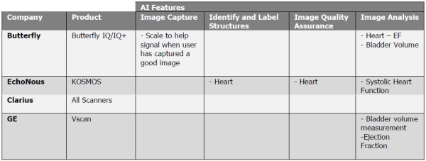
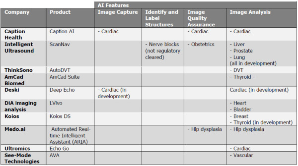
Ultrasound AI is at an early stage of development and there are few commercialised and regulatory-cleared solutions on the market. As shown in Tables 1 and 2, there are several use cases for AI, ranging from anatomy navigation and image capture assistance, to image quality assurance and image analysis. AI-powered anatomy navigation will help novice users to locate, identify and label parts of the anatomy, while image capture assistance will provide guidance on how best to position the probe to obtain the optimal image quality. The first solutions are coming to market, including Caption Health’s Caption AI which guides novice ultrasound users in acquiring diagnostic-quality echo exams, and Intelligent Ultrasound’s ScanNav Anatomy (CE and De Novo FDA applied for) which identifies several nerve types for peripheral nerve block procedures.
While AI for ultrasound image analysis has progressed faster, most of the products on the market today are “point” solutions for specific tasks, for example algorithmic solutions for automated assessment of ejection fraction of the heart or automated counting of B-lines for lung imaging. Moreover, these solutions are only available for the heart, lungs and thyroid and there are no AI-based image analysis products for other body areas, for example the liver, aorta and gallbladder. This limits the utility of ultrasound AI as it can only be applied to specific exams, whereas users require an AI “toolkit” with support for the most common exam and procedure types.
Going forward, today’s point solutions are expected to evolve into “comprehensive” AI solutions that can detect, label, quantify and classify multiple findings for a given organ or body area. Ultimately, comprehensive AI solutions will be available for multiple body areas and full body imaging. As AI begins to more closely mimic how clinicians use ultrasound, by offering image capture and diagnostic support for a wide range of common pathologies and findings, in addition to supporting with interventional procedures, the barriers to using ultrasound will be lowered and it will become more accessible to novice users.
AI-powered solutions for cardiac ultrasound are the most progressed, especially for image analysis, and closest to a comprehensive AI solution. For example, DiA Imaging Analysis markets a suite of tools including ejection fraction, strain and segmental wall motion analysis, but even in cardiac ultrasound the AI “toolbox” is far from complete. Ultimately, it could take several years before there are comprehensive AI solutions available for most routine exams. To bridge the gap until that time, novice users can be connected to ultrasound experts by teleultrasound platforms.
Teleultrasound — the bridge over troubled waters
Teleultrasound platforms can be used for every exam type and procedure, and in recent years platforms have been developed specifically for point of care ultrasound. These platforms allow for collaboration with specialist clinicians, which is particularly useful for novice users unfamiliar with ultrasound. It allows novice users to obtain ultrasound expertise, potentially from anywhere in the world, in real time, and the collaborator can interact with the novice user to assist with image capture to obtain quality images, and with the subsequent image interpretation. Teleultrasound is becoming increasingly important due to the lack of trained ultrasound users in some rural areas and developing countries.
The COVID-19 outbreak has been a recent driver, with teleultrasound platforms more in demand to connect clinicians in frontline care, many of whom are novice ultrasound users, with experts. The combination of teleultrasound and AI will enable ultrasound to not only be more portable, but to also be carried out more remotely.
The Signify View
Ultrasound was once touted as replacing the stethoscope; such was its promise of widespread use. While this has not happened, the prospect of it happening in the coming years is now more realistic. The ultrasound skills shortage is the last remaining barrier preventing handheld ultrasound from obtaining mainstream use in point of care and primary care settings. AI and teleultrasound platforms can help overcome this barrier and both trends are already underway and gaining pace. However, the AI solutions currently available have relatively limited clinical utility, and what is needed, and is absent at present, are comprehensive AI solutions. There has been a similar trend in other imaging modalities like CT and X-ray, where AI was initially applied to specific use cases, such as cancer detection. However, there are now AI packages for chest X-rays, for example, that can detect tens of radiological findings. It is this jump that is needed for ultrasound AI to take handheld ultrasound mainstream, but this will take time as new algorithms will need validation and FDA approval before going to market. Teleultrasound platforms are being used in the interim until these comprehensive AI solutions are ready. However even after this occurs, teleultrasound will still play a role in taking handhelds mainstream. They will remain a way of connecting users and will offer remote collaboration which AI alone cannot provide.
 About the author: Mustafa Hassan joined Signify Research in 2020 as part of the Medical Imaging team which covers areas such as ultrasound, general radiography and machine learning in medical imaging. Prior to that he obtained a Ph.D. in Pharmacy and Physiology from the University of Kent and has three years of post-doctoral experience working on optimising healthcare for genetic Cardiac diseases. In his spare time, he has a passion for sport and fitness and spending time with his wife and family.
Ultrasound scanners have streamlined over the years from being refrigerator-sized units in the 1980s to now being the size of a smartphone. These handheld ultrasound systems, aside from having the benefit of being ultraportable, are also cheaper than larger console units and are increasingly able to offer sufficient image quality and performance for most routine exams and procedures. An increasingly diverse range of handheld scanners are coming to market, with some targeted at experienced ultrasound users in specific clinical specialties and others designed for more general imaging applications.
Their ultra-portability, relatively low cost, and ease of disinfection mean that they are in high demand to combat the COVID-19 pandemic, with the handheld ultrasound market forecast to increase by around 70% worldwide in 2020. While proving popular to combat COVID-19, there are still limitations to the widespread utilisation of handheld ultrasound in routine clinical practice. Despite the market being forecast to nearly double in size from 2020 to 2024, handhelds are only forecast to account for around 6% of the total ultrasound market in 2024, with consoles and compacts still taking the lion’s share.
The barriers to mainstream use of handheld ultrasound are primarily cost, mindset, and education. The cost of handheld scanners has come down considerably in the last few years, to the extent that they are now at an affordable price point for most clinicians and physicians. The mindset has also improved, with medical students now learning how to use ultrasound during their training and thus, becoming more inclined to use the systems as they enter clinical practice. Moreover, the performance of handhelds has steadily improved over the years, both in terms of image quality and clinical utility. However, education remains a barrier, with many point-of-care clinicians lacking the technical ability to use handheld ultrasound. This is even more the case with primary care physicians. Two ways that technology can help overcome this barrier are with artificial intelligence (AI) and the use of teleultrasound.
AI –handheld ultrasound’s key enabler
AI is addressing the limitations of handheld ultrasound, and indeed, ultrasound in general, by helping the user to capture an ultrasound image, ensuring the quality of that image is sufficient and by assisting in the diagnostic analysis of the image. While ultrasound AI is still in the early stages of commercialisation, it will ultimately increase the number of new users of ultrasound by simplifying ease-of-use of the scanners.
Table 1 - AI solutions available for handheld ultrasounds
Ultrasound AI is at an early stage of development and there are few commercialised and regulatory-cleared solutions on the market. As shown in Tables 1 and 2, there are several use cases for AI, ranging from anatomy navigation and image capture assistance, to image quality assurance and image analysis. AI-powered anatomy navigation will help novice users to locate, identify and label parts of the anatomy, while image capture assistance will provide guidance on how best to position the probe to obtain the optimal image quality. The first solutions are coming to market, including Caption Health’s Caption AI which guides novice ultrasound users in acquiring diagnostic-quality echo exams, and Intelligent Ultrasound’s ScanNav Anatomy (CE and De Novo FDA applied for) which identifies several nerve types for peripheral nerve block procedures.
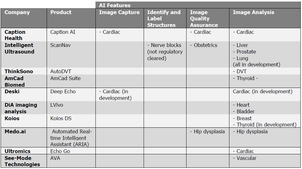
Table 2 - Solutions offered by ultrasound AI software developers
While AI for ultrasound image analysis has progressed faster, most of the products on the market today are “point” solutions for specific tasks, for example algorithmic solutions for automated assessment of ejection fraction of the heart or automated counting of B-lines for lung imaging. Moreover, these solutions are only available for the heart, lungs and thyroid and there are no AI-based image analysis products for other body areas, for example the liver, aorta and gallbladder. This limits the utility of ultrasound AI as it can only be applied to specific exams, whereas users require an AI “toolkit” with support for the most common exam and procedure types.
Going forward, today’s point solutions are expected to evolve into “comprehensive” AI solutions that can detect, label, quantify and classify multiple findings for a given organ or body area. Ultimately, comprehensive AI solutions will be available for multiple body areas and full body imaging. As AI begins to more closely mimic how clinicians use ultrasound, by offering image capture and diagnostic support for a wide range of common pathologies and findings, in addition to supporting with interventional procedures, the barriers to using ultrasound will be lowered and it will become more accessible to novice users.
AI-powered solutions for cardiac ultrasound are the most progressed, especially for image analysis, and closest to a comprehensive AI solution. For example, DiA Imaging Analysis markets a suite of tools including ejection fraction, strain and segmental wall motion analysis, but even in cardiac ultrasound the AI “toolbox” is far from complete. Ultimately, it could take several years before there are comprehensive AI solutions available for most routine exams. To bridge the gap until that time, novice users can be connected to ultrasound experts by teleultrasound platforms.
Teleultrasound — the bridge over troubled waters
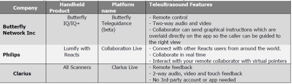
Teleultrasound platforms can be used for every exam type and procedure, and in recent years platforms have been developed specifically for point of care ultrasound. These platforms allow for collaboration with specialist clinicians, which is particularly useful for novice users unfamiliar with ultrasound. It allows novice users to obtain ultrasound expertise, potentially from anywhere in the world, in real time, and the collaborator can interact with the novice user to assist with image capture to obtain quality images, and with the subsequent image interpretation. Teleultrasound is becoming increasingly important due to the lack of trained ultrasound users in some rural areas and developing countries.
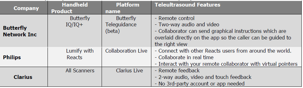
Table 3 - Examples of handheld ultrasound teleultrasound platforms
The COVID-19 outbreak has been a recent driver, with teleultrasound platforms more in demand to connect clinicians in frontline care, many of whom are novice ultrasound users, with experts. The combination of teleultrasound and AI will enable ultrasound to not only be more portable, but to also be carried out more remotely.
The Signify View
Ultrasound was once touted as replacing the stethoscope; such was its promise of widespread use. While this has not happened, the prospect of it happening in the coming years is now more realistic. The ultrasound skills shortage is the last remaining barrier preventing handheld ultrasound from obtaining mainstream use in point of care and primary care settings. AI and teleultrasound platforms can help overcome this barrier and both trends are already underway and gaining pace. However, the AI solutions currently available have relatively limited clinical utility, and what is needed, and is absent at present, are comprehensive AI solutions. There has been a similar trend in other imaging modalities like CT and X-ray, where AI was initially applied to specific use cases, such as cancer detection. However, there are now AI packages for chest X-rays, for example, that can detect tens of radiological findings. It is this jump that is needed for ultrasound AI to take handheld ultrasound mainstream, but this will take time as new algorithms will need validation and FDA approval before going to market. Teleultrasound platforms are being used in the interim until these comprehensive AI solutions are ready. However even after this occurs, teleultrasound will still play a role in taking handhelds mainstream. They will remain a way of connecting users and will offer remote collaboration which AI alone cannot provide.

Mustafa Hassan