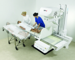
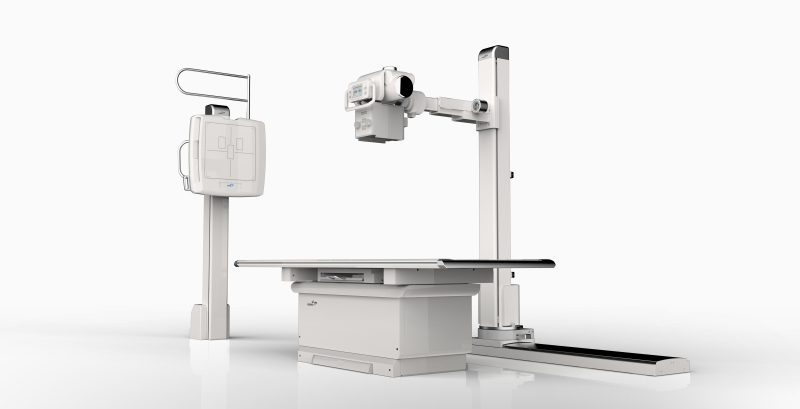
Agfa's DR 800 multipurpose X-ray room
Multifunctional and retrofitted: What’s new in radiography and fluoroscopy
November 21, 2017
by Lisa Chamoff, Contributing Reporter
As hospitals and imaging facilities prepare for cuts to Medicare reimbursements for exams performed using analog X-ray systems this year, and to exams using computed radiography (CR) equipment in 2018, manufacturers have been responding with DR retrofit solutions.
Medicare payments for analog X-rays have been cut by 20 percent as part of the Consolidated Appropriations Act of 2016. Starting next year, exams performed using CR equipment will be reduced by 7 percent for five years and will later be cut by 10 percent.
OEMs are also continuing to release all-in-one radiography and fluoroscopy systems to increase room utilization, with flexible positioning to increase patient comfort and image processing software designed to produce high-quality images with the lowest radiation dose possible.
Here are some of the new systems, software and retrofit solutions that have been released within the last year.
Agfa
Last year at RSNA, Agfa previewed its new multipurpose DR 800 X-ray room as a work in progress and expects to submit the 510(k) prior to this year's RSNA meeting.
The overhead tube system is the first product for Agfa that will be capable of doing general radiography, fluoroscopy and interventional studies. The company offers a predecessor to the system in Europe.
“It’s one of these systems that could be used all day long by radiologists and specialists outside radiology,” says George Curley, senior sales marketing manager for digital imaging at Agfa. “It really is a versatile room.”
The DR 800 will have a table that can go 90 degrees in either direction and an X-ray tube that can go out a full 180 centimeters.
“You can literally do chest X-rays on the table at full distance,” Curley says.
The system will also include Agfa’s MUSICA (Multi-Scale Image Contrast Amplification) image processing software and Dynamic MUSICA for fluoroscopy.
“MUSICA really is the best on the market for handling noise in low-dose images,” Curley says.
Carestream
Carestream recently introduced a new DirectView software option called SmartGrid.
"This enhancement algorithm automatically processes an image to create a scatter-reduced appearance,” says Sarah Verna, Carestream's marketing manager for global X-ray solutions. “It offers an important advantage since scatter introduces low frequencies into an image, which appears as a haze that reduces contrast and detail. The traditional means of reducing scatter is to use an anti-scatter grid, but this presents challenges to the technologist with positioning/alignment and increases dose to the patient.
"Many physical factors affect the properties of scatter: energy spectrum of the beam; thickness/size of the object and collimation. Using empirical modeling, SmartGrid can accommodate these factors through estimation of the algorithm parameters which are tuned to approximate anti-scatter grid visual performance.”
Fujifilm
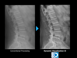
Fujifilm recently released its Dynamic Visualization II image processing software, a new generation that uses artificial intelligence to adjust image quality based on structural recognition
“The previous generation of Dynamic Visualization dynamically enhanced various areas of the image for contrast adjustment,” says Rob Fabrizio, director of marketing and product development for digital X-ray at FUJIFILM Medical Systems U.S.A., Inc. “It didn’t have the automatic recognition in terms of structural densities, the shape and characteristic of bones and orthopedic hardware. It’s really a dramatic difference when you see the before and after images, especially on low-penetration and low-dose X-rays.”
Dynamic Visualization II is available for Fujifilm’s mini portable X-ray system and the company will be rolling it out for its FDR Go mobile and digital radiography rooms and retrofits.
“There’s a lot of intelligence built into it from our years of digital medical image processing experience,” Fabrizio says. “Orthopedic hardware won’t have a halo effect around the edges. All areas will be enhanced regardless of varying densities.”
GE Healthcare
Since RSNA last year, GE Healthcare has launched multiple new offerings.
“All of them are aimed at helping our customers make the first image count,” says Gokhan Gunes, X-ray marketing director for GE Healthcare.
The company's new X-ray Quality Application features Repeat Reject Analytics and is part of the Applied Intelligence platform (it was in beta at RSNA 2016). It connects to compatible new and legacy X-ray systems, and uses web-based dashboards to manage quality assurance, helping customers uncover the root causes of rejected X-rays so the department can deploy improvements.
"Before, manually collecting repeat/reject data was very cumbersome and time-consuming. You had to go from system to system across multiple sites to aggregate data," Gunes says.
The tool was built in collaboration with the University of Washington and Humber River Hospital in Canada.
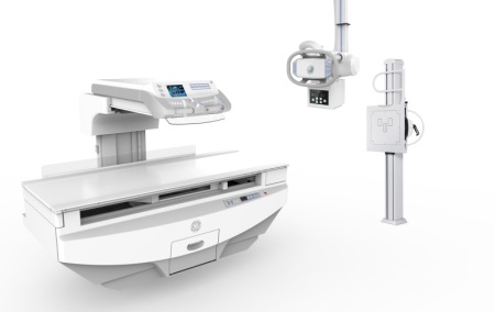
Also unveiled at RSNA last year was the Proteus XR/f, GE Healthcare's new floor-mounted digital radiography system, which Gunes says provides a more simplified transition to digital for outpatient facilities. The system, which can be used for all exam types and fits in smaller rooms, comes with multiple-size wireless digital detectors, and auto tracking, a form of automation that Gunes says is generally found in ceiling-mounted rooms.
"It's a system that packs a lot of flexibility and clinical capability into a compact unit," Gunes says. "We made sure that we were able to offer a smooth transition to DR with little disruption in practice."
The Precision 600FP is a premium flat-panel digital fluoroscopy system also unveiled at RSNA 2016. Similar to the Proteus XR/f, the Precision 600FP is designed for all patient types and can fit in fluoroscopy rooms with limited space. It also comes with a comprehensive dose management package.
The system is geared toward facilities that have a need for a dedicated fluoroscopy room and have a diverse patient population.
“There was demand here,” Gunes says. “We’re looking at facilities that require a dedicated room and want to provide a low-dose fluoroscopy exam to a wide variety of patients without going through construction.”
Konica Minolta
At last year’s RSNA, Konica Minolta released its KDR Advanced U-Arm (AU), a small footprint X-ray system programmed by exam anatomy.
“When you walk in with a patient, all you have to do is adjust it up and down a little and you’re ready to go,” says Guillermo Sander, DR senior marketing manager for Konica Minolta. “Customers have said it has improved their workflow.”
The best use for the system is orthopedic imaging, Sander says, and some hospitals are using it in their children’s orthopedic wing.
Sander says the system’s “slick design” also makes the machine look less intimidating to patients.
“We’ve learned that patients are more comfortable when the machine doesn’t look as intimidating,” Sander says. “We spent a lot of time … improving the patient experience.”
Novarad
This summer, Novarad released its Chameleon DR, the first modality for the enterprise imaging and workflow solutions company.
The DR retrofit system utilizes the company’s proprietary post-processing software, which Steve Fowler, Novarad’s director of product management, says makes the Chameleon DR stand out from other retrofit solutions.
The algorithms in the image processing software take patient size and density into account.
“The resulting images are nothing short of amazing,” Fowler says. “Physicians and technicians are seeing extremely well-balanced bone-to-tissue displays that enable more confident diagnoses.”
The 100-micron detector, in combination with the image processing software, means exams require less radiation dose, Fowler says, while the Chameleon’s multi-procedure workflow allows technologists to position the patient less often, which leads to less painful, faster exams.
The system is geared toward imaging centers and hospitals seeking a budget-friendly upgrade to DR, with reimbursement changes around the corner.
“There are some very inexpensive solutions out there, but their pixel pitch and resolution is a definite compromise,” Fowler says. “For some facilities that are tight on budget, it will at least get them compliant with the new reimbursement mandate. The Chameleon is for those facilities that are more forward-thinking and acquiring a platform that will deliver lower doses, faster reads and better health outcomes.”
Philips
This year, Philips introduced the CombiDiagnost R90, a remote controlled digital radiography and fluoroscopy system with dose management technology. The system, which received FDA approval in late January, is built for high room utilization and throughput, says Fabrice Moulene, senior product manager for diagnostic X-Ray and fluoroscopy at Philips.
Philips’ Dynamic UNIQUE image processing software allows images to be taken at lower doses, and uses what Moulene says is a real-time, frame-by-frame de-noising, “so images are cleaned from noise from the first frame onwards, so radiologists don’t need to wait.”
Combined with a movement compensation between frames, the software also completely removes ghosting artifacts and has an automatic, real-time brightness adjustment, resulting in a very stable brightness of the whole acquired sequence, Moulene says.
“With this technology, we are seeing significantly improved image quality, as well as higher diagnostic confidence among radiologists,” Moulene says. “Dynamic UNIQUE not only sets a new benchmark in the market for high quality fluoroscopy at low dose, but also the advanced image processing that can benefit the workflow and clinical outcome in a customer’s room. It harmonizes contrast and enhances faint details to provide consistent image quality for both radiography and fluoroscopy images, and provides de-noising capabilities to enhance image quality especially in low dose imaging.”
The CombiDiagnost R90 is geared toward medium to large hospitals with insufficient workload in their fluoroscopy rooms, looking for increased efficiency and flexibility. While Philips found that its customers’ fluoroscopy rooms were underutilized, as the number of exams were on the decline, they still required a fluoroscopy system.
“CombiDiagnost R90 was developed to address this exact challenge,” Moulene says. “With this technology, users can perform both digital radiography and fluoroscopy procedures in one room, optimizing equipment while reducing costs and the number of needed procedure rooms.”

Rayence
At last year’s RSNA, Rayence debuted its XR-5 Digital Radiography System, a floor-mounted DR system that is well positioned for orthopedic practices, imaging centers and urgent care centers.
The system, which has an auto-tracking feature, can be installed in a room with eight-foot ceilings and requires less power — 208 to 220 volts — than traditional X-ray systems and comes at a competitive price, says Bill Nicholas, director of corporate marketing and communications at Rayence.
Shimadzu
Last year, Shimadzu introduced the T-Smart technology to its Sonialvision G4 all-in-one DR and fluoroscopy system. T-Smart is an optional workstation with enhanced tomosynthesis for general radiography, not just for breast imaging.
“It takes the great images of tomosynthesis and reprocesses them with new algorithms and makes them even sharper,” says Frank Serrao, marketing manager for Shimadzu. “It also reduces the blooming effect of metal artifacts.”
Serrao says the enhanced data can show hairline fractures that often can’t be seen with CT or MR.
“When we do tomosynthesis on a G4 … we’re allowed to be able to put the patient on the table and stand the table at 90 degrees,” Serrao says. “When you’re lying down, there’s no weight bearing, so you can’t see anything. You have to image it in weight-bearing conditions.”
At this year’s RSNA, Shimadzu is introducing a new fixed radiography system that is in the process of being cleared. The company is waiting until the process is further along to release more information.

Siemens Healthineers
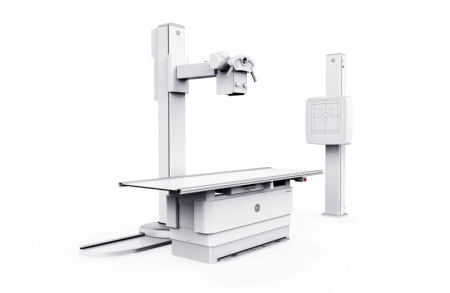
At the end of 2016, Siemens Healthineers released the Multix Fusion Max, its mid-range, ceiling-mounted X-ray room that is geared toward general radiography for small- and medium-sized hospitals as well as outpatient practices. The system has the option of a bone stitching package for facilities doing occasional orthopedic imaging.
Customers can also easily swap wireless detectors with other systems that are part of the Max family, including the Mobilett Mira Max and Ysio Max, increasing uptime, says Joseph D’Antonio, director of marketing for radiography, fluoroscopy, mobile and twin robotic X-ray products at Siemens Healthineers.
“The ability to swap detectors makes it good for standardizing departments and replacing an entire fleet of radiography and portable systems,” D’Antonio says.
The user interface is also the same within the Max family, adding to ease of use.
“Seventy-five percent of the components used in the Fusion Max are the same used in the Ysio Max,” D’Antonio says.
Joe Kidd, an X-ray technologist at the Soin Family Practice Center in Beavercreek, Ohio, uses the Multix Fusion Max and says he appreciates the system’s ease of use and speed.
“I think I’ve only had to repeat one image because the system is so easy to use,” Kidd says. “You also have the ability to see the image within six to nine seconds. It pops on your screen sometimes before you’re ready to look at it. With the first digital machine that I used, it took up to a minute to see the image.”
Siemens has also begun to install the 3-D option for the Multitom Rax Twin Robotic X-ray scanner, which was FDA cleared at the end of 2015 and has twin robotic arms that move around the patient during exams.
The 3-D option allows for the ability to image while the patient is in a natural weight-bearing position.
“Structures are going to show up much differently in a natural weight-bearing way,” D’Antonio says.
The Multitom Rax is also a good multipurpose option for facilities.
“Customers are very excited about the multi-use aspect,” D’Antonio says. “You can move swallow studies, angio studies and routine rad exams through the Multitom Rax very quickly. It’s allowed our customers to better use their assets and to combine systems. Instead of adding a fluoro room they add the Multitom Rax.”
Since not all customers are ready to move to a fully digital platform, Siemens introduced its DR retrofit, ArtPix EZ2GO, at the end of last year. It uses a detector from the same family as the Fusion Max and Ysio Max, so customers that eventually decide to fully upgrade don’t need to purchase another detector, splitting the upgrade into two phases.
“We’ve been interested in the retrofit market for a while,” D’Antonio says. “With the looming reimbursement changes, we have seen a high level of interest. Clearly, other vendors have been in the retrofit market for years, but our ability to provide an on-ramp for digital and an on-ramp for Siemens technology makes us unique.”
Medicare payments for analog X-rays have been cut by 20 percent as part of the Consolidated Appropriations Act of 2016. Starting next year, exams performed using CR equipment will be reduced by 7 percent for five years and will later be cut by 10 percent.
OEMs are also continuing to release all-in-one radiography and fluoroscopy systems to increase room utilization, with flexible positioning to increase patient comfort and image processing software designed to produce high-quality images with the lowest radiation dose possible.
Here are some of the new systems, software and retrofit solutions that have been released within the last year.
Agfa
Last year at RSNA, Agfa previewed its new multipurpose DR 800 X-ray room as a work in progress and expects to submit the 510(k) prior to this year's RSNA meeting.
The overhead tube system is the first product for Agfa that will be capable of doing general radiography, fluoroscopy and interventional studies. The company offers a predecessor to the system in Europe.
“It’s one of these systems that could be used all day long by radiologists and specialists outside radiology,” says George Curley, senior sales marketing manager for digital imaging at Agfa. “It really is a versatile room.”
The DR 800 will have a table that can go 90 degrees in either direction and an X-ray tube that can go out a full 180 centimeters.
“You can literally do chest X-rays on the table at full distance,” Curley says.
The system will also include Agfa’s MUSICA (Multi-Scale Image Contrast Amplification) image processing software and Dynamic MUSICA for fluoroscopy.
“MUSICA really is the best on the market for handling noise in low-dose images,” Curley says.
Carestream
Carestream recently introduced a new DirectView software option called SmartGrid.
"This enhancement algorithm automatically processes an image to create a scatter-reduced appearance,” says Sarah Verna, Carestream's marketing manager for global X-ray solutions. “It offers an important advantage since scatter introduces low frequencies into an image, which appears as a haze that reduces contrast and detail. The traditional means of reducing scatter is to use an anti-scatter grid, but this presents challenges to the technologist with positioning/alignment and increases dose to the patient.
"Many physical factors affect the properties of scatter: energy spectrum of the beam; thickness/size of the object and collimation. Using empirical modeling, SmartGrid can accommodate these factors through estimation of the algorithm parameters which are tuned to approximate anti-scatter grid visual performance.”

Fujifilm's Dynamic Visualization II
image processing software
image processing software
Fujifilm
Fujifilm recently released its Dynamic Visualization II image processing software, a new generation that uses artificial intelligence to adjust image quality based on structural recognition
“The previous generation of Dynamic Visualization dynamically enhanced various areas of the image for contrast adjustment,” says Rob Fabrizio, director of marketing and product development for digital X-ray at FUJIFILM Medical Systems U.S.A., Inc. “It didn’t have the automatic recognition in terms of structural densities, the shape and characteristic of bones and orthopedic hardware. It’s really a dramatic difference when you see the before and after images, especially on low-penetration and low-dose X-rays.”
Dynamic Visualization II is available for Fujifilm’s mini portable X-ray system and the company will be rolling it out for its FDR Go mobile and digital radiography rooms and retrofits.
“There’s a lot of intelligence built into it from our years of digital medical image processing experience,” Fabrizio says. “Orthopedic hardware won’t have a halo effect around the edges. All areas will be enhanced regardless of varying densities.”

GE Healthcare's Precision 600FP premium flat-panel digital fluoroscopy system
GE Healthcare
Since RSNA last year, GE Healthcare has launched multiple new offerings.
“All of them are aimed at helping our customers make the first image count,” says Gokhan Gunes, X-ray marketing director for GE Healthcare.
The company's new X-ray Quality Application features Repeat Reject Analytics and is part of the Applied Intelligence platform (it was in beta at RSNA 2016). It connects to compatible new and legacy X-ray systems, and uses web-based dashboards to manage quality assurance, helping customers uncover the root causes of rejected X-rays so the department can deploy improvements.
"Before, manually collecting repeat/reject data was very cumbersome and time-consuming. You had to go from system to system across multiple sites to aggregate data," Gunes says.
The tool was built in collaboration with the University of Washington and Humber River Hospital in Canada.
The Proteus XR/f, GE Healthcare's new floor-mounted digital radiography system
Also unveiled at RSNA last year was the Proteus XR/f, GE Healthcare's new floor-mounted digital radiography system, which Gunes says provides a more simplified transition to digital for outpatient facilities. The system, which can be used for all exam types and fits in smaller rooms, comes with multiple-size wireless digital detectors, and auto tracking, a form of automation that Gunes says is generally found in ceiling-mounted rooms.
"It's a system that packs a lot of flexibility and clinical capability into a compact unit," Gunes says. "We made sure that we were able to offer a smooth transition to DR with little disruption in practice."
The Precision 600FP is a premium flat-panel digital fluoroscopy system also unveiled at RSNA 2016. Similar to the Proteus XR/f, the Precision 600FP is designed for all patient types and can fit in fluoroscopy rooms with limited space. It also comes with a comprehensive dose management package.
The system is geared toward facilities that have a need for a dedicated fluoroscopy room and have a diverse patient population.
“There was demand here,” Gunes says. “We’re looking at facilities that require a dedicated room and want to provide a low-dose fluoroscopy exam to a wide variety of patients without going through construction.”
Konica Minolta
At last year’s RSNA, Konica Minolta released its KDR Advanced U-Arm (AU), a small footprint X-ray system programmed by exam anatomy.
“When you walk in with a patient, all you have to do is adjust it up and down a little and you’re ready to go,” says Guillermo Sander, DR senior marketing manager for Konica Minolta. “Customers have said it has improved their workflow.”
The best use for the system is orthopedic imaging, Sander says, and some hospitals are using it in their children’s orthopedic wing.
Sander says the system’s “slick design” also makes the machine look less intimidating to patients.
“We’ve learned that patients are more comfortable when the machine doesn’t look as intimidating,” Sander says. “We spent a lot of time … improving the patient experience.”

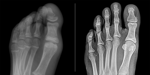
Before (left) and after (right) image processing
on the Novarad Chameleon DR retrofit system
on the Novarad Chameleon DR retrofit system
Novarad
This summer, Novarad released its Chameleon DR, the first modality for the enterprise imaging and workflow solutions company.
The DR retrofit system utilizes the company’s proprietary post-processing software, which Steve Fowler, Novarad’s director of product management, says makes the Chameleon DR stand out from other retrofit solutions.
The algorithms in the image processing software take patient size and density into account.
“The resulting images are nothing short of amazing,” Fowler says. “Physicians and technicians are seeing extremely well-balanced bone-to-tissue displays that enable more confident diagnoses.”
The 100-micron detector, in combination with the image processing software, means exams require less radiation dose, Fowler says, while the Chameleon’s multi-procedure workflow allows technologists to position the patient less often, which leads to less painful, faster exams.
The system is geared toward imaging centers and hospitals seeking a budget-friendly upgrade to DR, with reimbursement changes around the corner.
“There are some very inexpensive solutions out there, but their pixel pitch and resolution is a definite compromise,” Fowler says. “For some facilities that are tight on budget, it will at least get them compliant with the new reimbursement mandate. The Chameleon is for those facilities that are more forward-thinking and acquiring a platform that will deliver lower doses, faster reads and better health outcomes.”

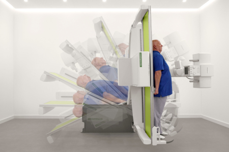
The Philips CombiDiagnost R90 digital radiography and fluoroscopy system
Philips
This year, Philips introduced the CombiDiagnost R90, a remote controlled digital radiography and fluoroscopy system with dose management technology. The system, which received FDA approval in late January, is built for high room utilization and throughput, says Fabrice Moulene, senior product manager for diagnostic X-Ray and fluoroscopy at Philips.
Philips’ Dynamic UNIQUE image processing software allows images to be taken at lower doses, and uses what Moulene says is a real-time, frame-by-frame de-noising, “so images are cleaned from noise from the first frame onwards, so radiologists don’t need to wait.”
Combined with a movement compensation between frames, the software also completely removes ghosting artifacts and has an automatic, real-time brightness adjustment, resulting in a very stable brightness of the whole acquired sequence, Moulene says.
“With this technology, we are seeing significantly improved image quality, as well as higher diagnostic confidence among radiologists,” Moulene says. “Dynamic UNIQUE not only sets a new benchmark in the market for high quality fluoroscopy at low dose, but also the advanced image processing that can benefit the workflow and clinical outcome in a customer’s room. It harmonizes contrast and enhances faint details to provide consistent image quality for both radiography and fluoroscopy images, and provides de-noising capabilities to enhance image quality especially in low dose imaging.”
The CombiDiagnost R90 is geared toward medium to large hospitals with insufficient workload in their fluoroscopy rooms, looking for increased efficiency and flexibility. While Philips found that its customers’ fluoroscopy rooms were underutilized, as the number of exams were on the decline, they still required a fluoroscopy system.
“CombiDiagnost R90 was developed to address this exact challenge,” Moulene says. “With this technology, users can perform both digital radiography and fluoroscopy procedures in one room, optimizing equipment while reducing costs and the number of needed procedure rooms.”
Rayence's XR-5 Digital Radiography System
Rayence
At last year’s RSNA, Rayence debuted its XR-5 Digital Radiography System, a floor-mounted DR system that is well positioned for orthopedic practices, imaging centers and urgent care centers.
The system, which has an auto-tracking feature, can be installed in a room with eight-foot ceilings and requires less power — 208 to 220 volts — than traditional X-ray systems and comes at a competitive price, says Bill Nicholas, director of corporate marketing and communications at Rayence.

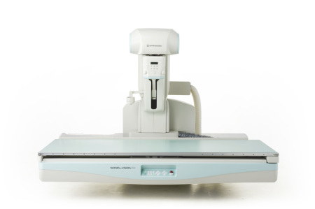
Shimadzu's new T-Smart technology is available on its Sonialvision G4 DR and fluoroscopy system
Shimadzu
Last year, Shimadzu introduced the T-Smart technology to its Sonialvision G4 all-in-one DR and fluoroscopy system. T-Smart is an optional workstation with enhanced tomosynthesis for general radiography, not just for breast imaging.
“It takes the great images of tomosynthesis and reprocesses them with new algorithms and makes them even sharper,” says Frank Serrao, marketing manager for Shimadzu. “It also reduces the blooming effect of metal artifacts.”
Serrao says the enhanced data can show hairline fractures that often can’t be seen with CT or MR.
“When we do tomosynthesis on a G4 … we’re allowed to be able to put the patient on the table and stand the table at 90 degrees,” Serrao says. “When you’re lying down, there’s no weight bearing, so you can’t see anything. You have to image it in weight-bearing conditions.”
At this year’s RSNA, Shimadzu is introducing a new fixed radiography system that is in the process of being cleared. The company is waiting until the process is further along to release more information.

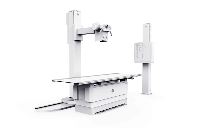
Siemens Healthineers' Multix Fusion Max X-ray system
Siemens Healthineers
At the end of 2016, Siemens Healthineers released the Multix Fusion Max, its mid-range, ceiling-mounted X-ray room that is geared toward general radiography for small- and medium-sized hospitals as well as outpatient practices. The system has the option of a bone stitching package for facilities doing occasional orthopedic imaging.
Customers can also easily swap wireless detectors with other systems that are part of the Max family, including the Mobilett Mira Max and Ysio Max, increasing uptime, says Joseph D’Antonio, director of marketing for radiography, fluoroscopy, mobile and twin robotic X-ray products at Siemens Healthineers.
“The ability to swap detectors makes it good for standardizing departments and replacing an entire fleet of radiography and portable systems,” D’Antonio says.
The user interface is also the same within the Max family, adding to ease of use.
“Seventy-five percent of the components used in the Fusion Max are the same used in the Ysio Max,” D’Antonio says.
Joe Kidd, an X-ray technologist at the Soin Family Practice Center in Beavercreek, Ohio, uses the Multix Fusion Max and says he appreciates the system’s ease of use and speed.
“I think I’ve only had to repeat one image because the system is so easy to use,” Kidd says. “You also have the ability to see the image within six to nine seconds. It pops on your screen sometimes before you’re ready to look at it. With the first digital machine that I used, it took up to a minute to see the image.”
Siemens has also begun to install the 3-D option for the Multitom Rax Twin Robotic X-ray scanner, which was FDA cleared at the end of 2015 and has twin robotic arms that move around the patient during exams.
The 3-D option allows for the ability to image while the patient is in a natural weight-bearing position.
“Structures are going to show up much differently in a natural weight-bearing way,” D’Antonio says.
The Multitom Rax is also a good multipurpose option for facilities.
“Customers are very excited about the multi-use aspect,” D’Antonio says. “You can move swallow studies, angio studies and routine rad exams through the Multitom Rax very quickly. It’s allowed our customers to better use their assets and to combine systems. Instead of adding a fluoro room they add the Multitom Rax.”
Since not all customers are ready to move to a fully digital platform, Siemens introduced its DR retrofit, ArtPix EZ2GO, at the end of last year. It uses a detector from the same family as the Fusion Max and Ysio Max, so customers that eventually decide to fully upgrade don’t need to purchase another detector, splitting the upgrade into two phases.
“We’ve been interested in the retrofit market for a while,” D’Antonio says. “With the looming reimbursement changes, we have seen a high level of interest. Clearly, other vendors have been in the retrofit market for years, but our ability to provide an on-ramp for digital and an on-ramp for Siemens technology makes us unique.”